

Trace of their ancestors can be dated from 485.4 million to 443.8 million years, which is why they are sometimes referred to as living fossils.
Best known species is the Limulus polyphemus, an American species which can grow to 60 centimeters in size and has a set of the largest compound eyes amongst arthropods. The total number of eyes is 10, which is already interesting by itself, as the eyes are located at different parts of the horseshoe crab’s body. A precise description can be found in the Encyclopedia of the Eye.
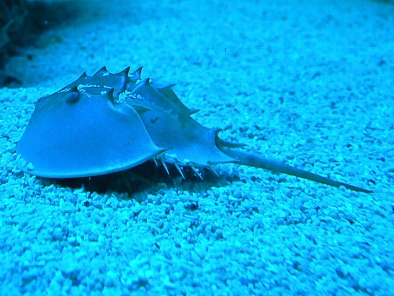
“American horseshoe crabs (Limulus polyphemus) have 10 eyes. They have two large lateral compound eyes, each containing about 1000 clusters of photoreceptors or ommatidia. A small lens within each ommatidium focuses light from a small patch of visual space onto each photoreceptor cluster, which transmits information about local changes in light intensity to the brain through a nerve fiber. There are five additional eyes on the top side of its first (anterior) major body section: two median eyes, one endoparietal eye, and two rudimentary lateral eyes. Two ventral eyes are located on the underside of the animal above the mouth. Photoreceptors located on the telson (tail) constitute the 10th eye.” [R. Payne, Y. Wang, in Encyclopedia of the Eye, 2010.]
Q-Phase microscope was used as one of the methods for analysis of the horseshoe crab’s cornea and its optical performance. The study of living creatures and cells composing their matter, is what allows the human kind to continuously explore the unseen, we are glad to take part in this ongoing process of discovery.
Gradients of Orientation, Composition, and Hydration of Proteins for Efficient Light Collection by the Cornea ofthe Horseshoe Crab
Article by O. Spaeker, et al. 2022
Abstract:
The lateral eyes of the horseshoe crab, Limulus polyphemus, are the largest compound eyes within recent Arthropoda. While this visual system has been extensively described before, the precise mechanism allowing vision has remained controversial. Correlating quantitative refractive index (RI) mapping and detailed structural analysis, we demonstrate how gradients of RI in the cornea result from the hierarchical organization of chitin-protein fibers, heterogeneity in protein composition and bromine doping, as well as spatial variation in water content. Combining the realistic cornea structure and measured RI gradients with full-wave optical modelling and ray-tracing approaches, we show that the light collection mechanism depends on both refraction-based graded index (GRIN) optics and total internal reflection. The optical properties of the cornea are governed by different mechanisms at different hierarchical levels, demonstrating the remarkable versatility of arthropod cuticle.