

Stem cells
Studying stem cell types and their differentiation properties helps to understand how they mature into functionally specialized cells.
Stem cell applications hold great promise as potential replacement therapy in regenerative medicine and other health-related problems such as spinal injuries, type-1 diabetes, Alzheimer’s disease, burns, and cancer.
The two defining characteristics of a stem cell are perpetual self-renewal and the ability to differentiate into a specialized adult cell type.
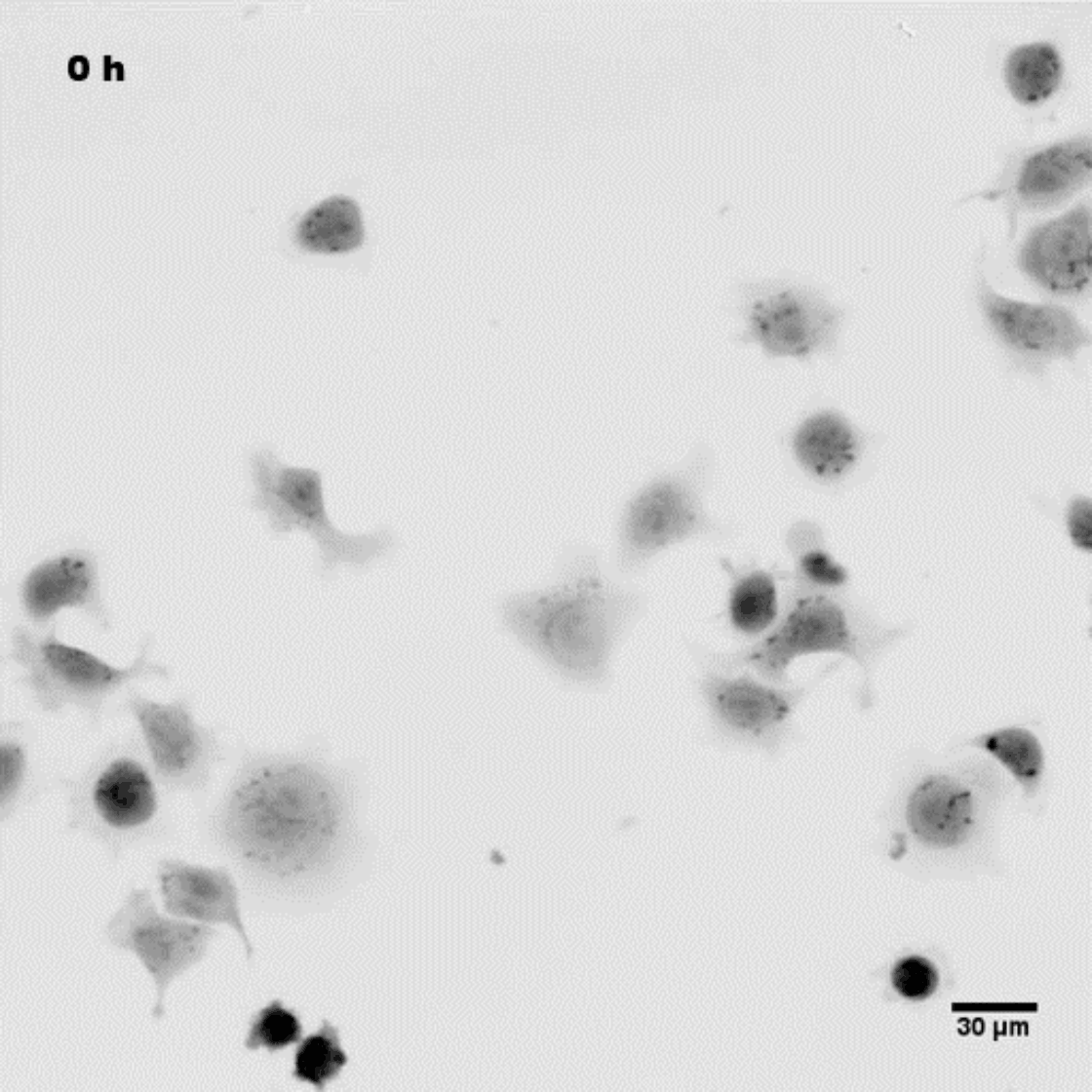
Q-Phase technology provides high phase detection sensitivity, allowing for measuring the cell dry mass of divided cells in pg/µm2 and thus defines cells with the morphological asymmetric division.
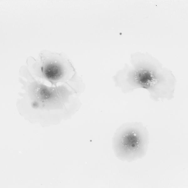
In addition, the clarity of the produced images ensures a precise segmentation and identification of delicate cell processes for detailed morphological assessment during differentiation. Q-Phase fluorescent module works simultaneously with QPI to visualize the differentiation markers.

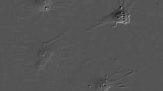
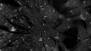
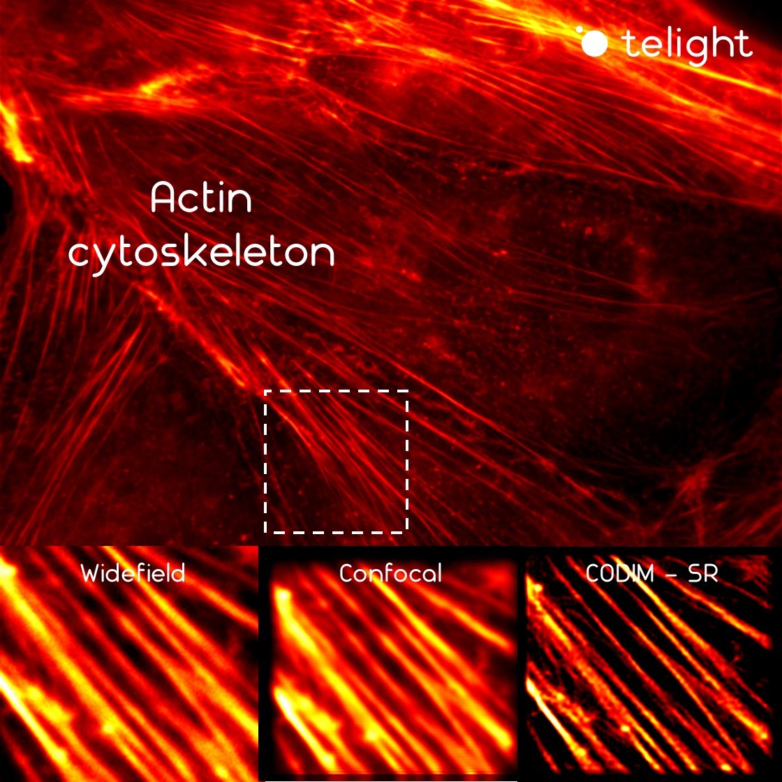
Due to the fundamental function of stem cells in developmental biology and their potential use in regenerative medicine and rare genetic disease treatment, the visualization of their differentiation gained high importance. LiveCodim super-resolution module enables the investigation of fundamental mechanisms driving stem cells on the molecular level.
Publications
Garita-Hernandez M., et al.
Optogenetic light sensors in retinal organoids
Products
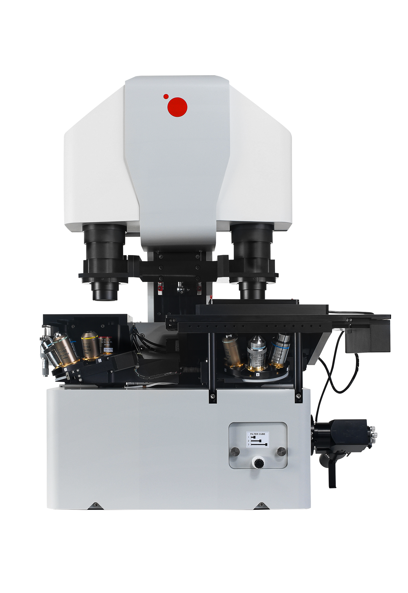
Q-Phase
Quantitative Phase Imaging
Q-Phase is a patented holographic microscope with high detection sensitivity, designed for gentle live-cell imaging.
Q-Phase is an ideal solution for experts who desire precise automated segmentation of individual cells for subsequent data analysis. Q-Phase quickly transforms cell features and dynamics into numerical data suitable for comparisons, correlations, and more detailed statistics.
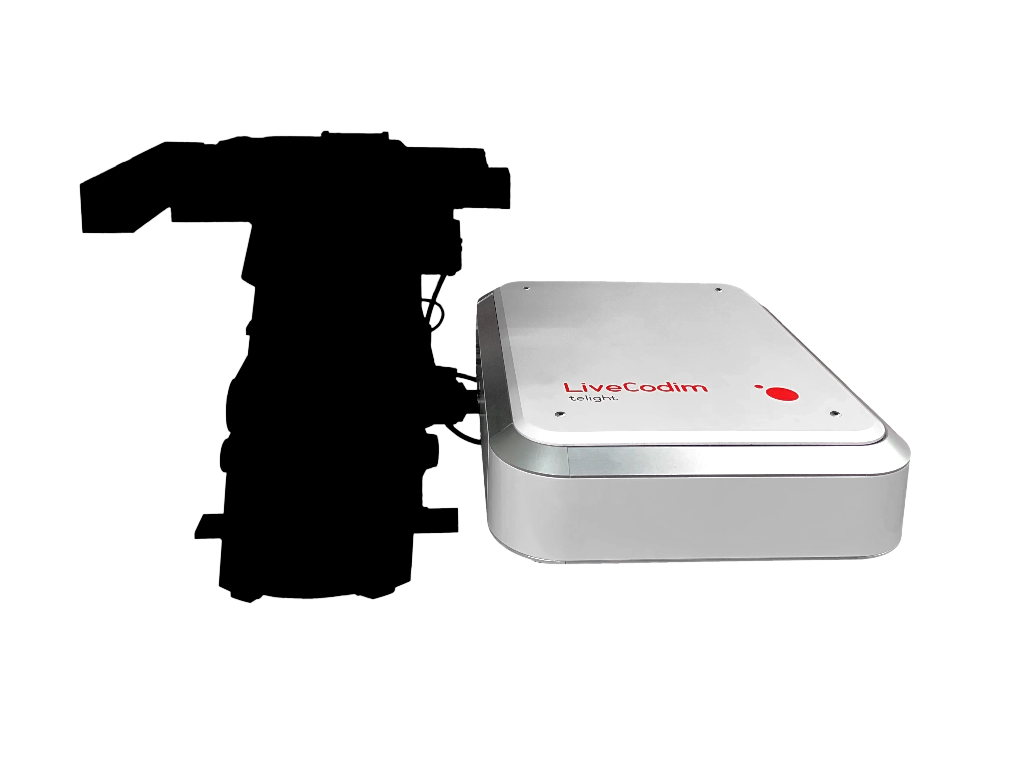
LiveCodim
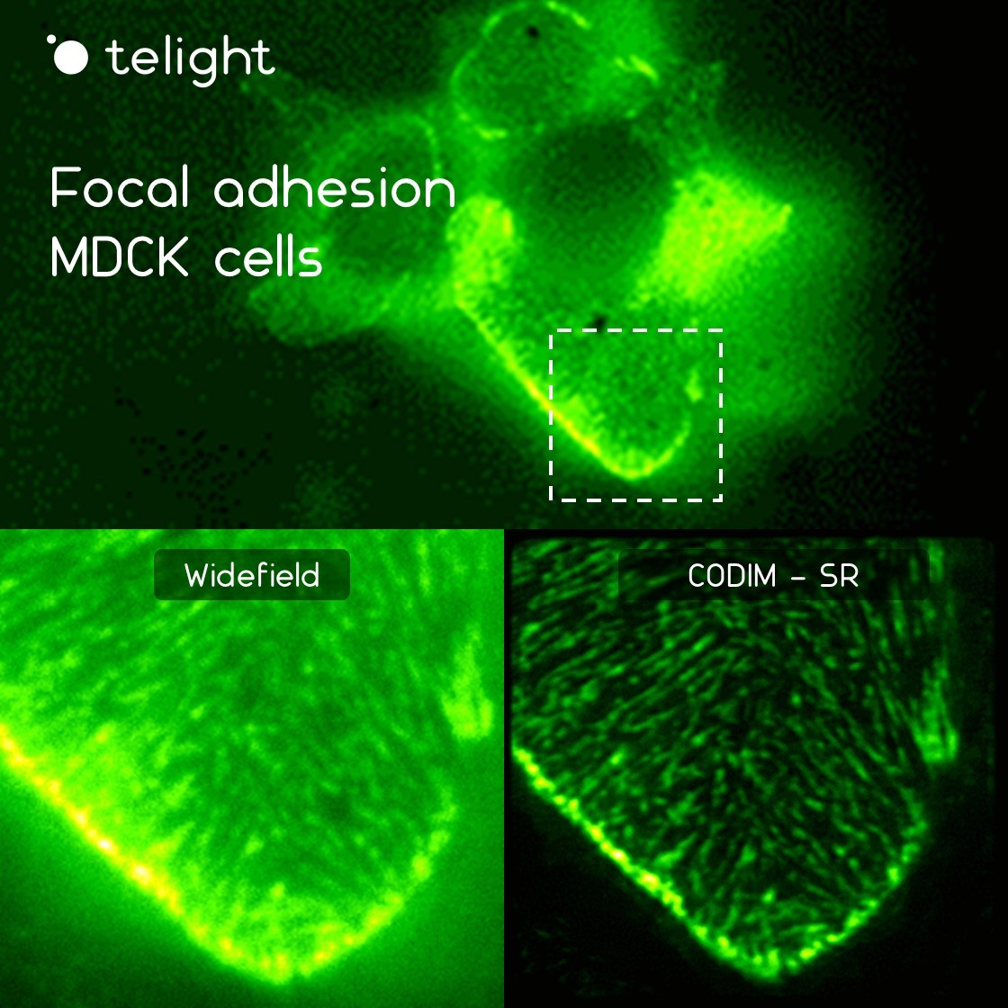
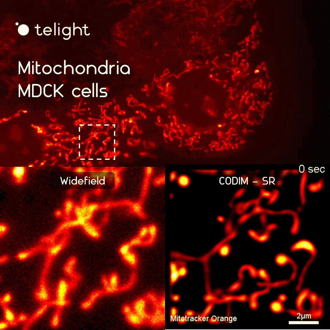
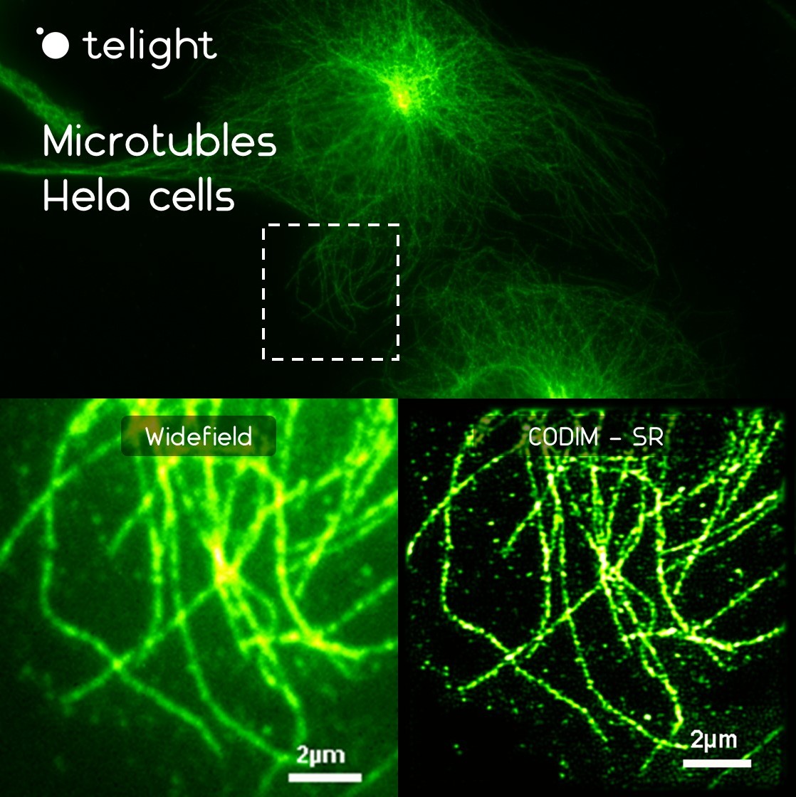
From conventional to super-resolution microscopy
LiveCodim is a universal, super-resolution imaging platform designed to interface with any standard fluorescence microscope. It is the solution for live-cell imaging with high resolution and low phototoxicity.