

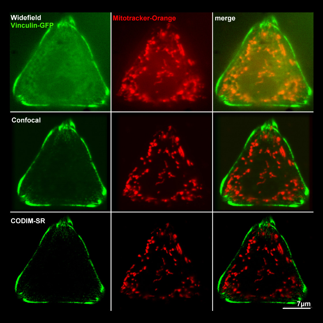
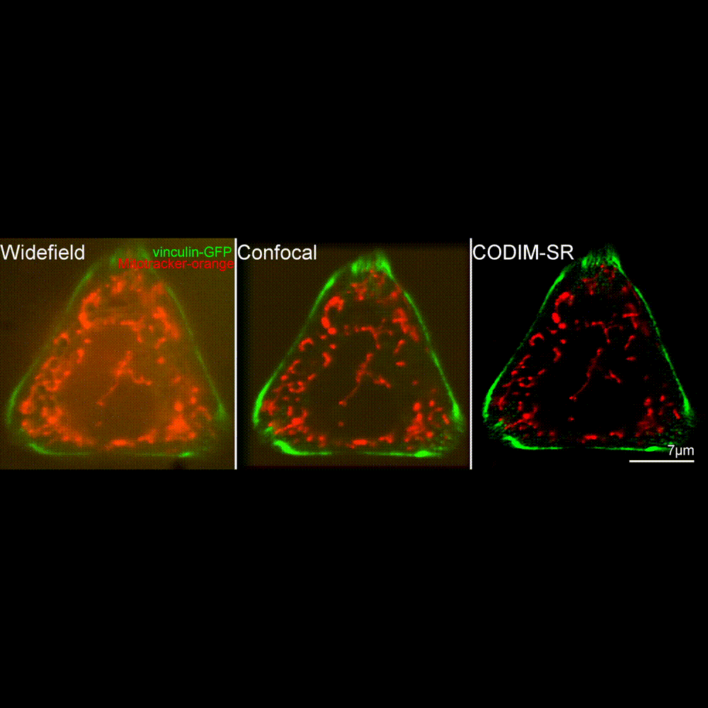
Super-resolution microscopy
With the development of new techniques, fluorescence optical microscopy can now help biologists decipher the dynamics of biological processes at the scale of a few tenths of a nanometer.
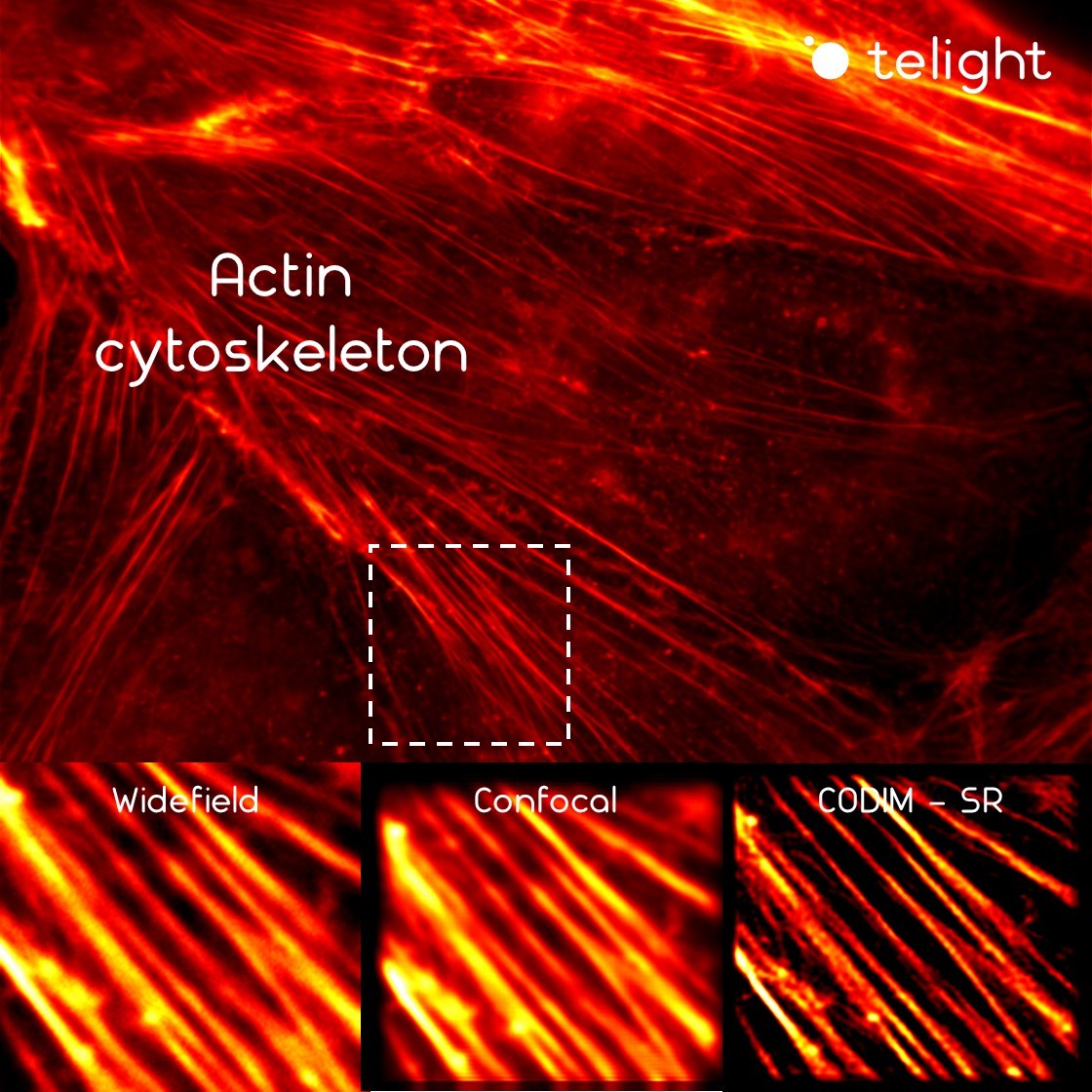
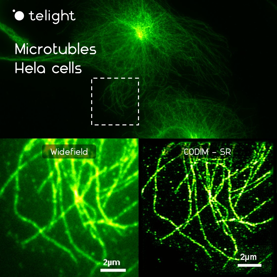
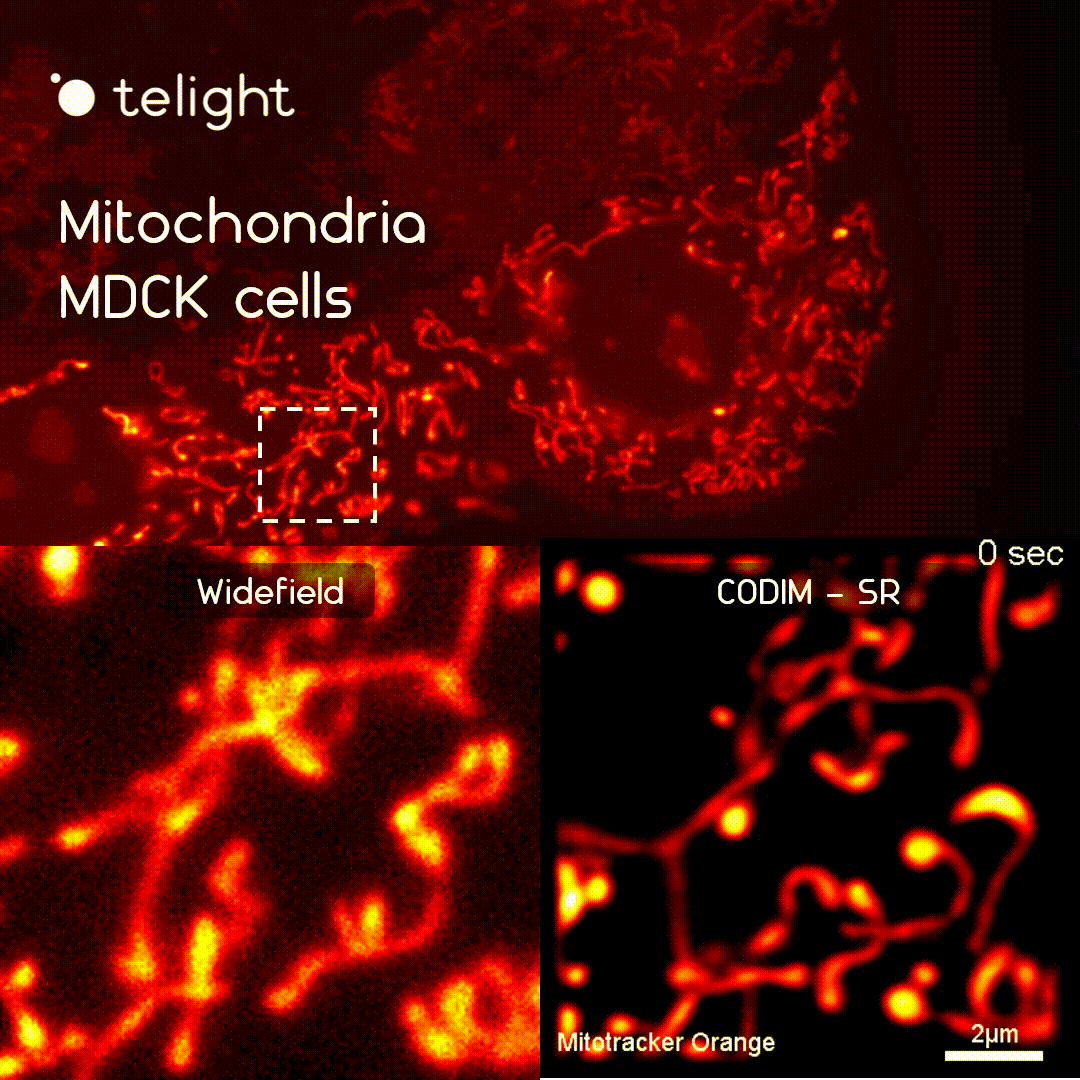
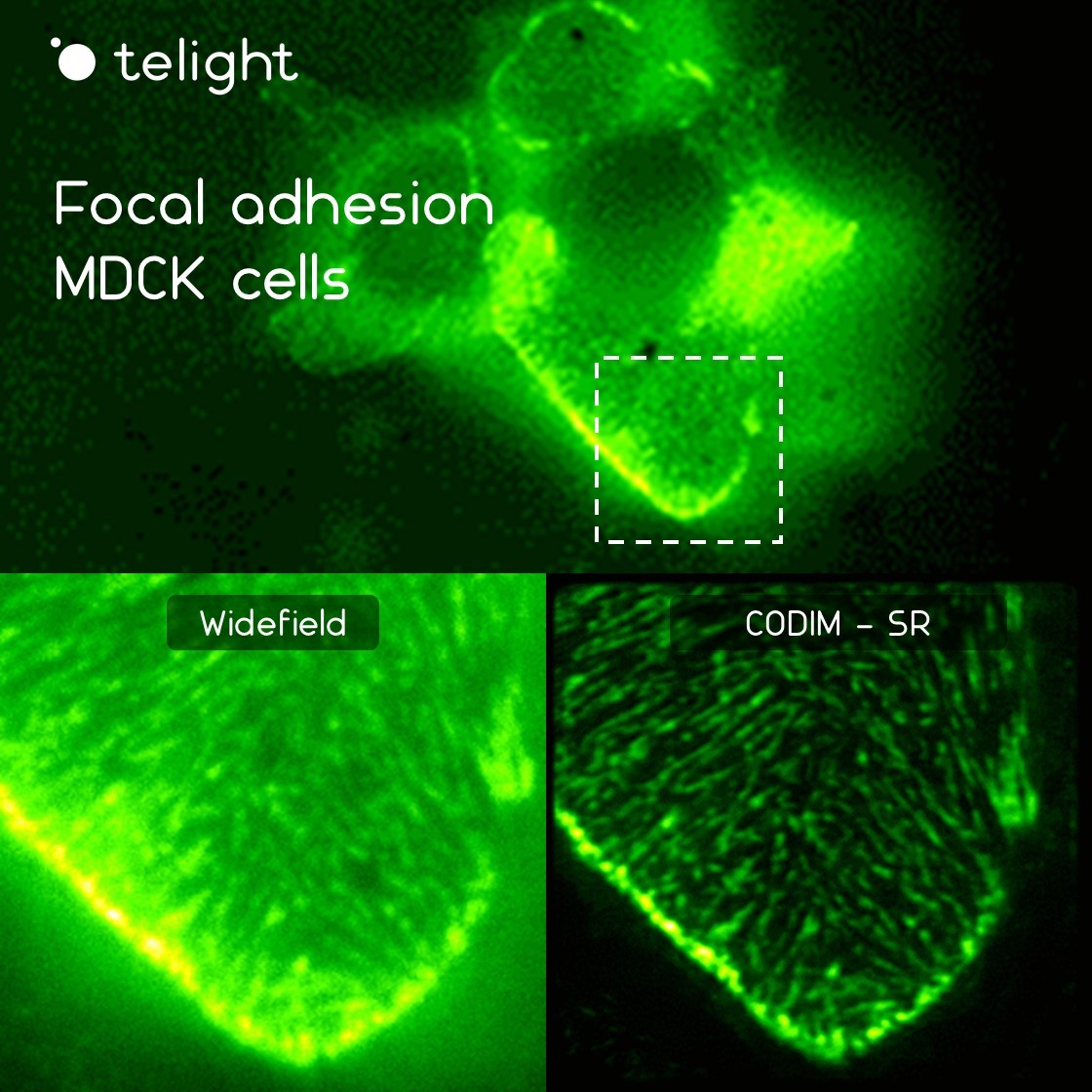
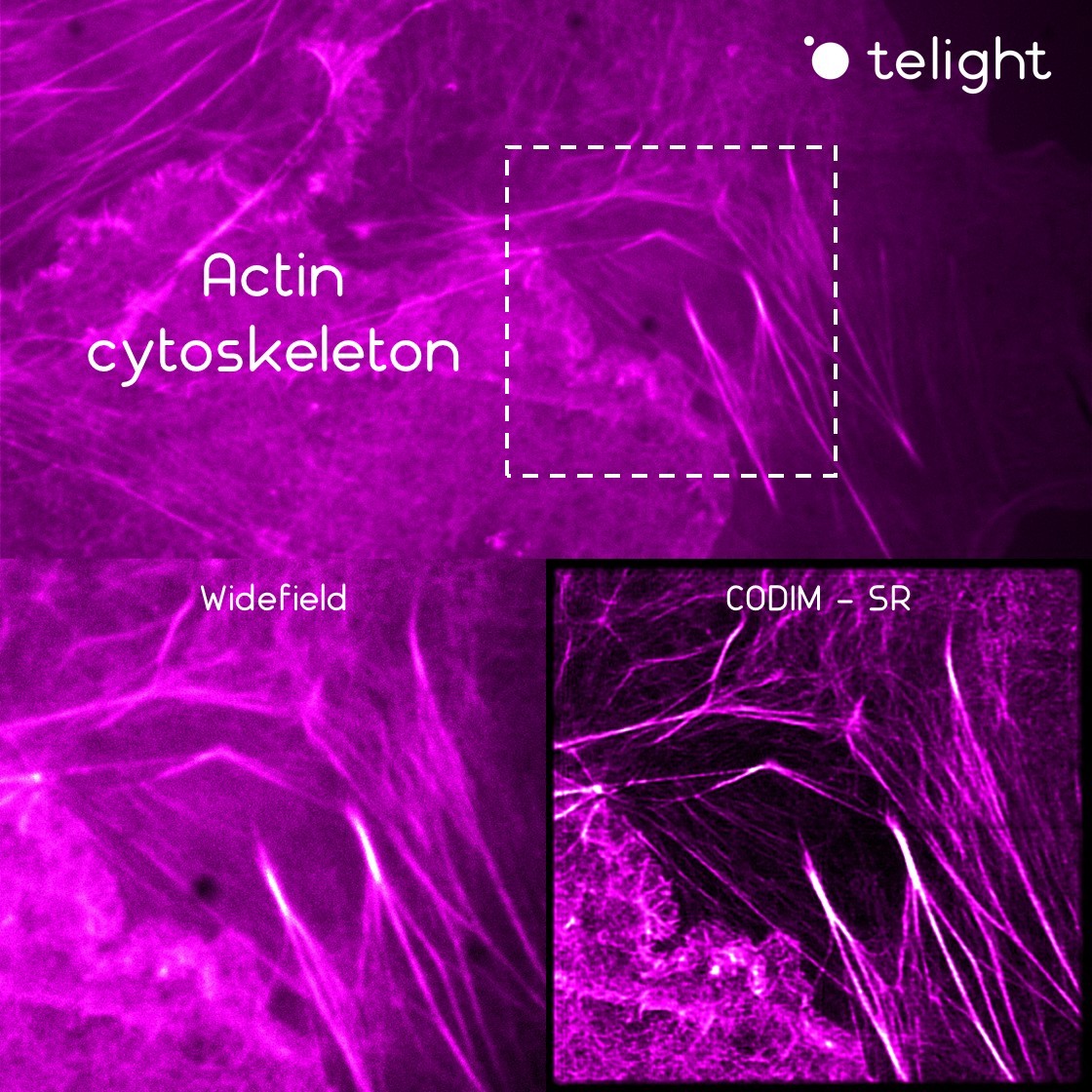
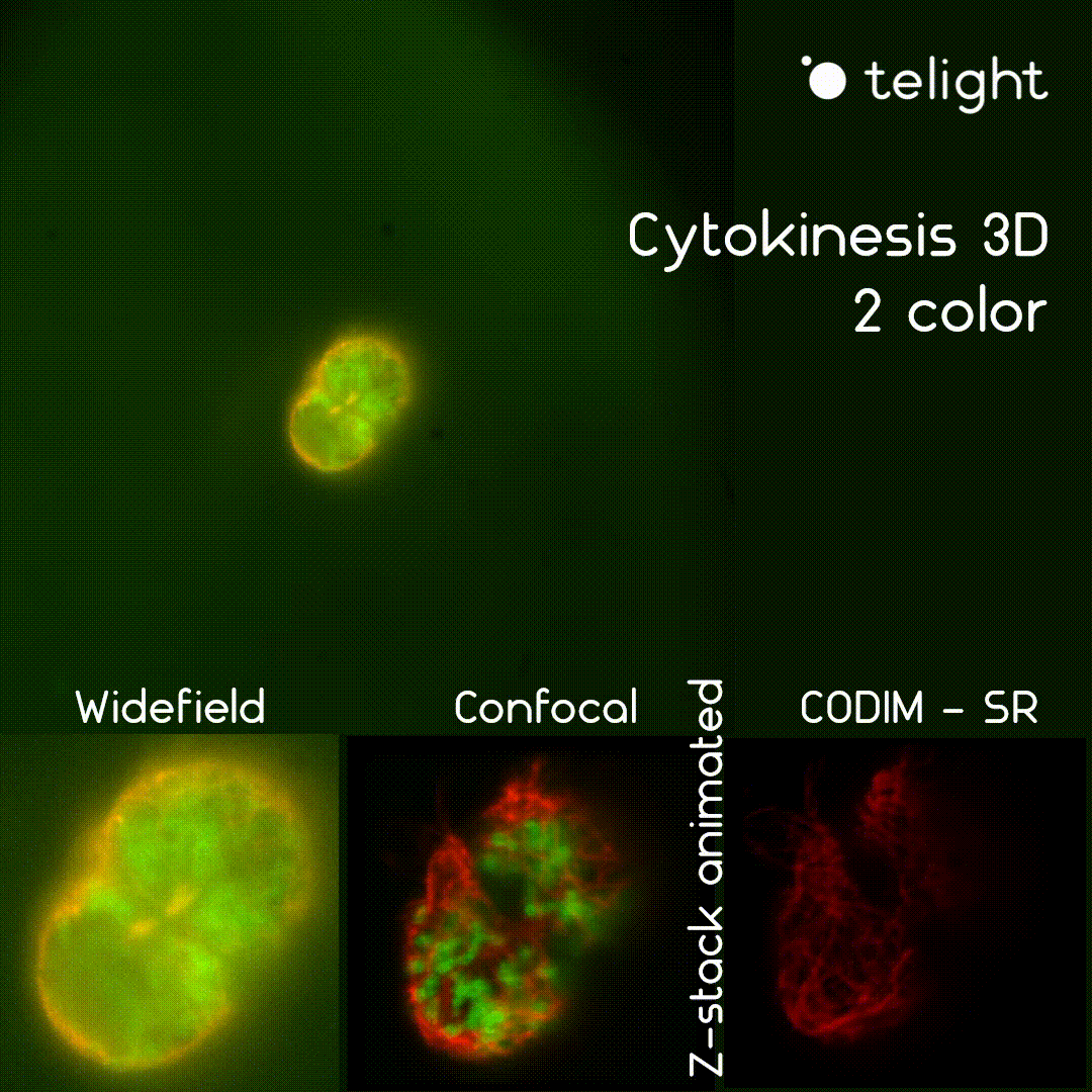
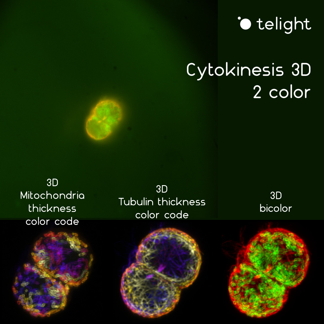
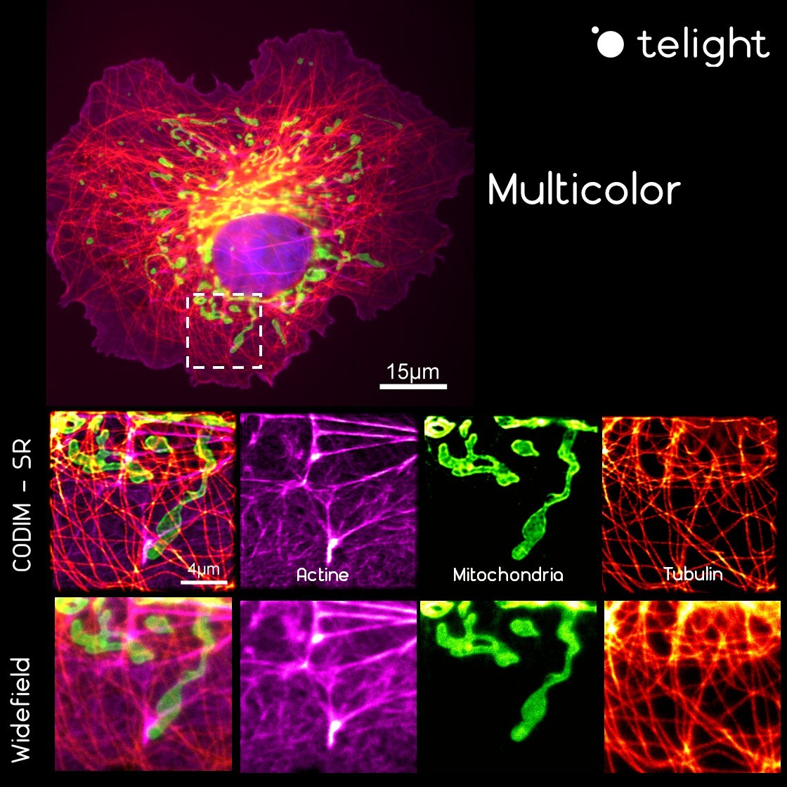
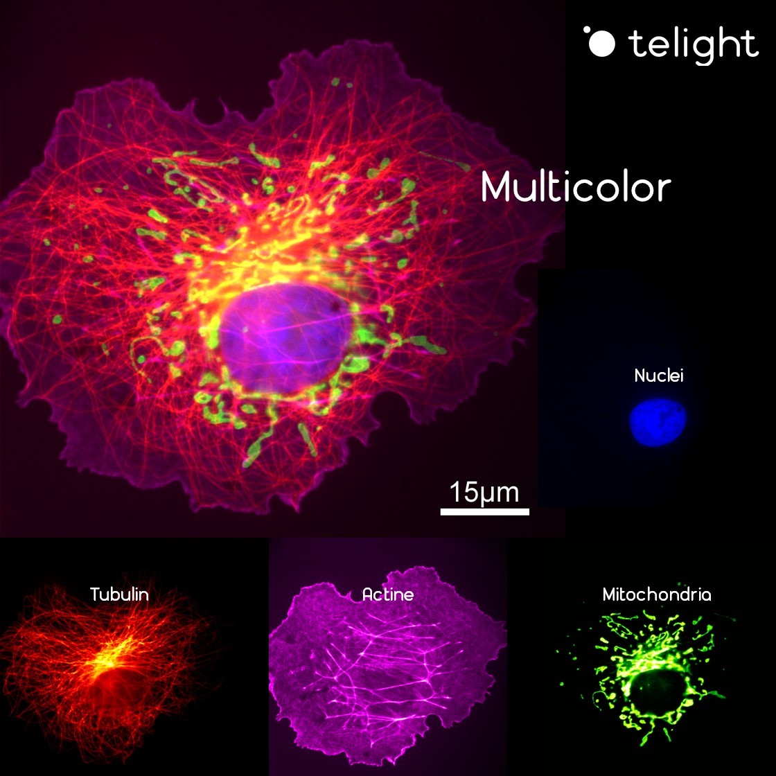
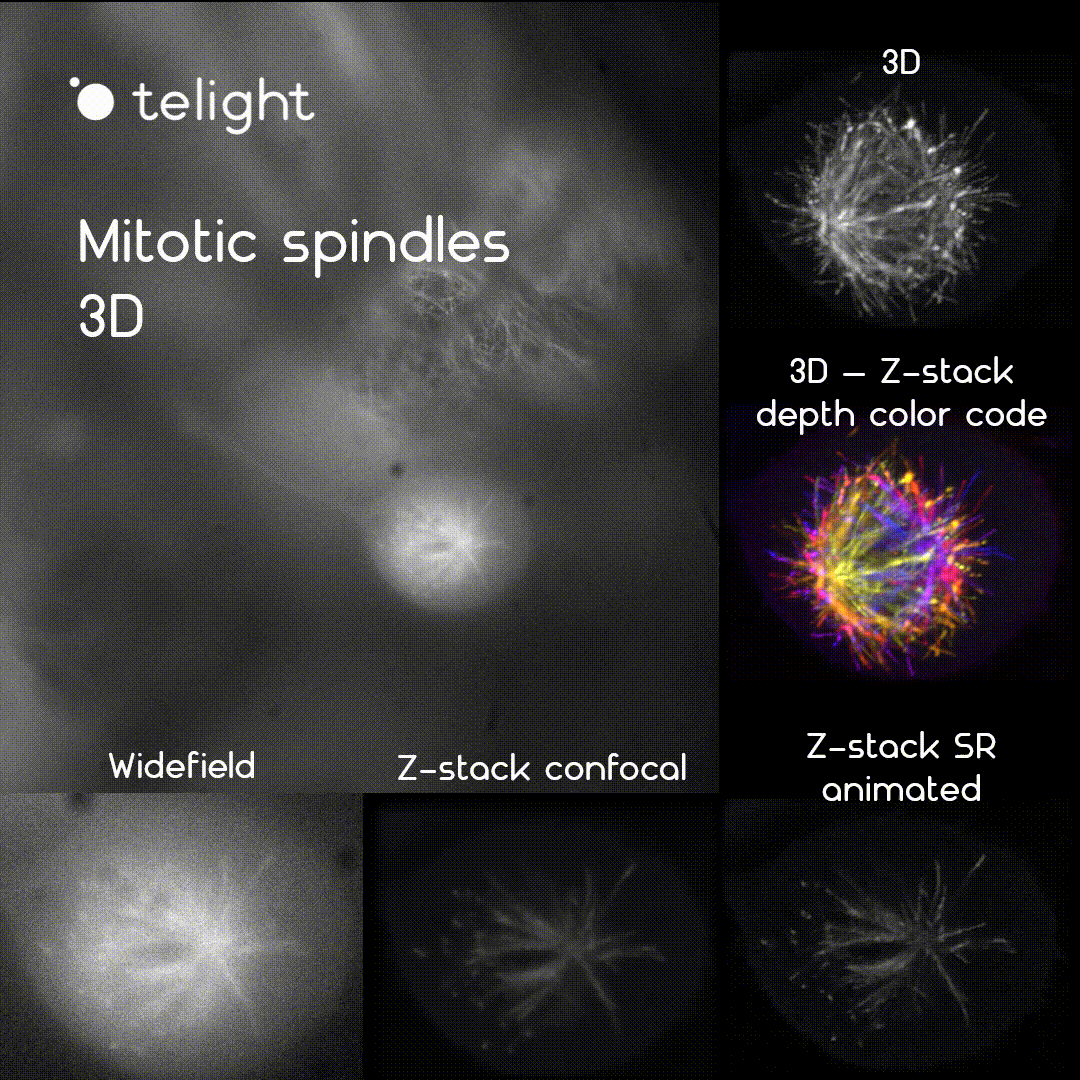
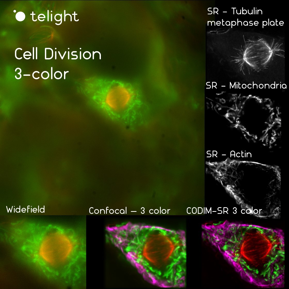
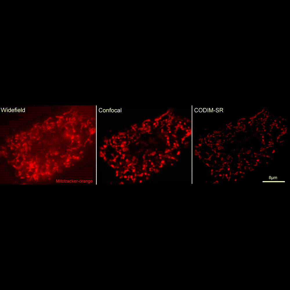
LiveCodim uses a unique patented laser beam shaper. Our technology perfectly fits the needs of biologists in super-resolution and is adapted for living samples, to visualize cellular and molecular dynamics in high resolution.
Publications
J. Vargas, et al.
The Wnt/Ca2+ pathway is involved in interneuronal communication mediated by tunneling nanotubes
Maarifi, G., Fernandez, J., Portilho, D.M., et al.
RanBP2 regulates the anti-retroviral activity of TRIM5α by SUMOylation at a predicted phosphorylated SUMOylation motif
Garita-Hernandez M., et al.
Optogenetic light sensors in retinal organoids
Getz A.M., Xu F., Visser F., et al.
Tumor suppressor menin is required for subunit-specific nAChR Alpha5 transcription and nAChR-dependent presynaptic facilitation in cultured mouse hippocampal neurons
Products
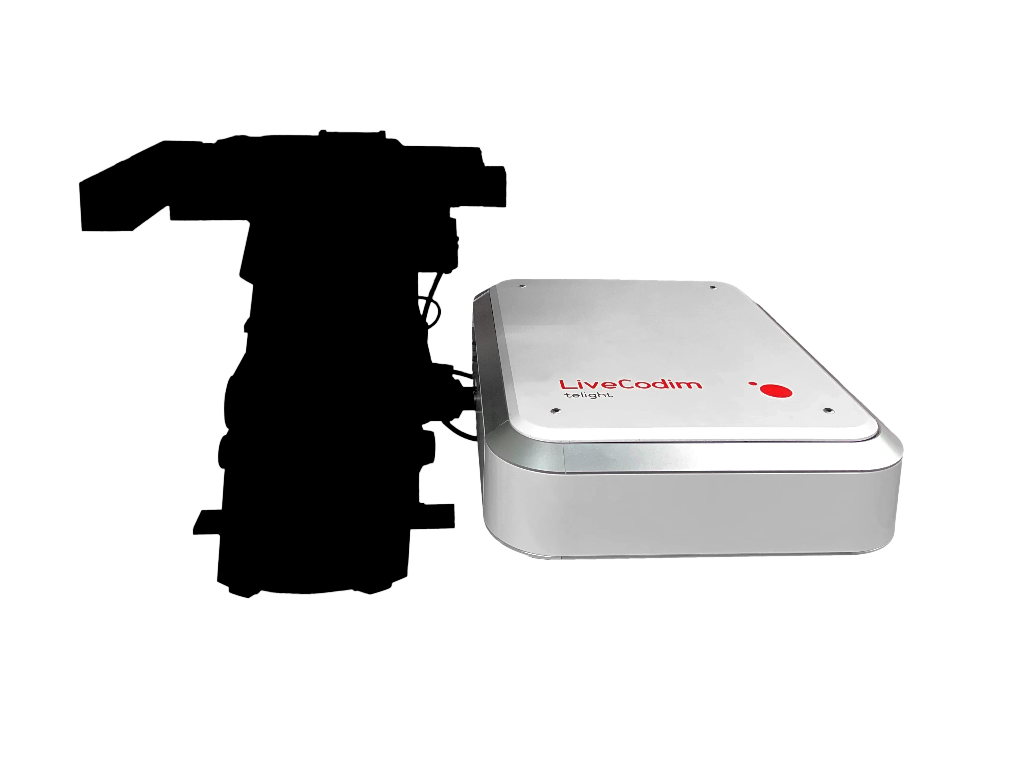
LiveCodim
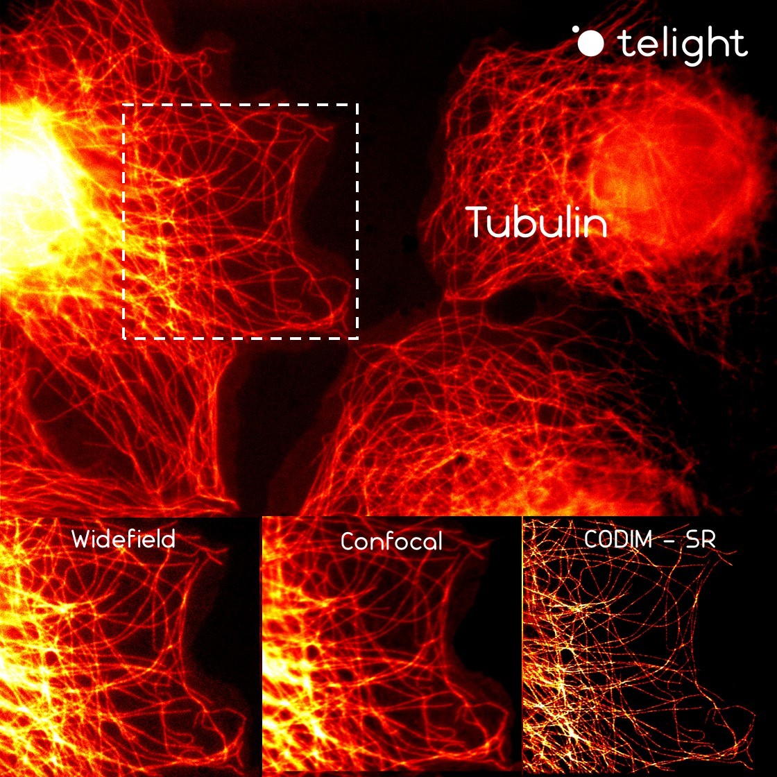
From conventional to super-resolution microscopy
LiveCodim is a universal, super-resolution imaging platform designed to interface with any standard fluorescence microscope. It is the solution for live-cell imaging with high resolution and low phototoxicity.