

Virology
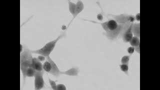
A virus is a submicroscopic structure containing genetic material that can not replicate without the machinery and metabolism of host cells. Viral infection is usually associated with morphological and pathological changes of infected cells that are necessary for efficient viral replication and may result in host cell death.
Time-lapse microscopy systems are essential for understanding the key factors influencing the viral replication steps and their effect on host cells.
Viral cytopathic effects on cell morphology are commonly observed as rounding of the infected cell, fusion with adjacent cells (syncytia), and the appearance as nuclear or cytoplasmic inclusion bodies. The virus can also alter the behavior of the host cells, suc as the cell cycle.
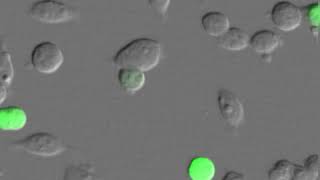
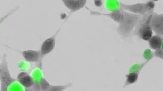
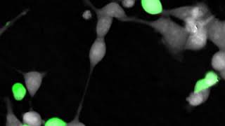
Q-Phase can monitor the influence of the virus on cells, mode of cell death, and immune response, all of that in real-time. The possibility to use both QPI and fluorescence in a single experiment enables to probe of the viral particles and simultaneously detect their development inside cells.
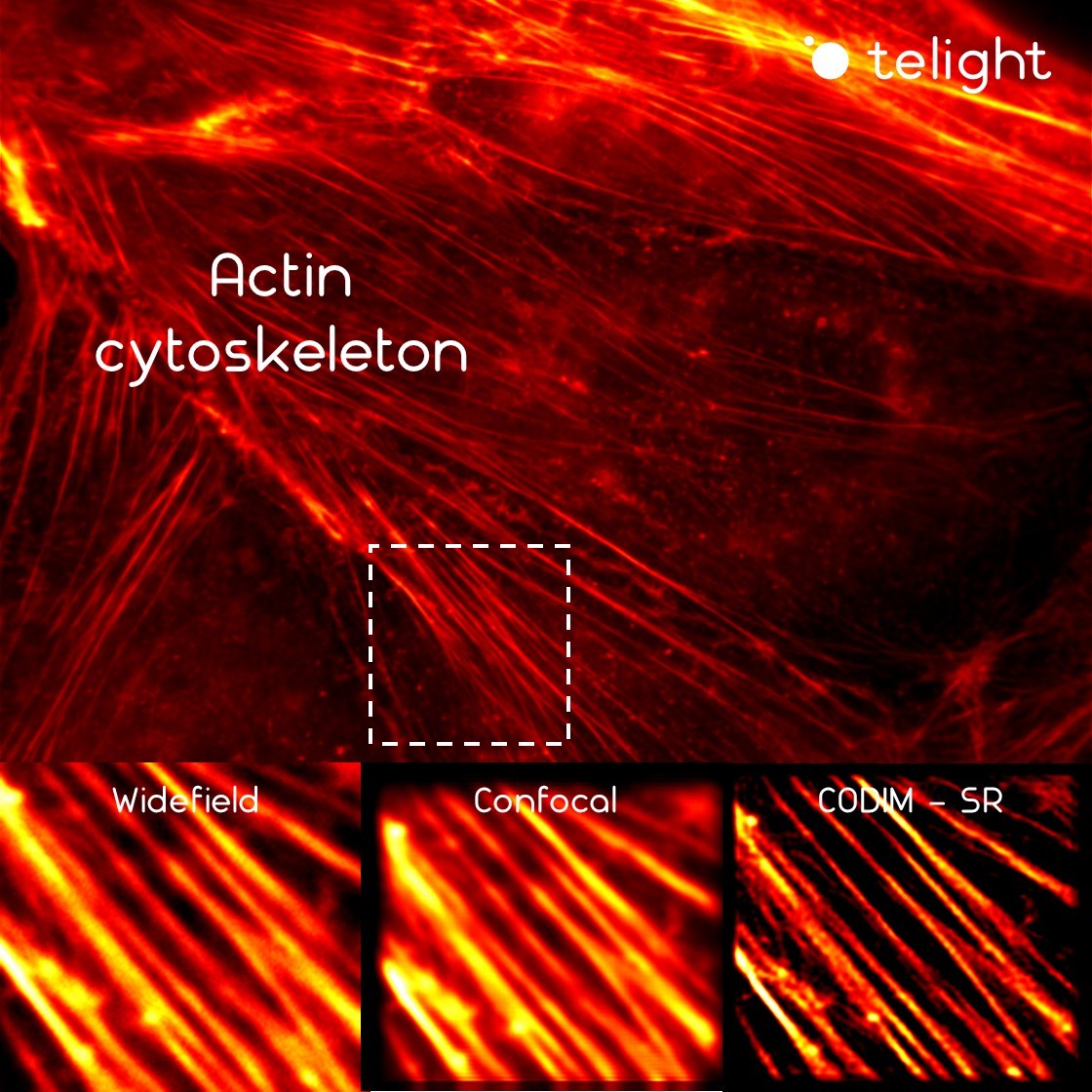
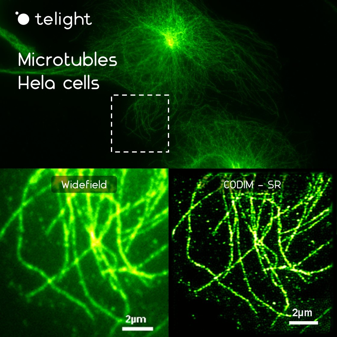
Due to their small size ranging between 20-200 nm, imaging of viruses using optical microscopy has been challenging for decades.
In this context, super-resolution techniques including LiveCodim will be of interest. It can provide visualization of different steps of cellular infection such as membrane interaction, transfer of genetic material, viral replication.
Publications
Broad-Spectrum Antiviral Activity of 3’-Deoxy-3’-Fluoroadenosine against Emerging Flaviviruses
Maarifi, G., Fernandez, J., Portilho, D.M., et al.
RanBP2 regulates the anti-retroviral activity of TRIM5α by SUMOylation at a predicted phosphorylated SUMOylation motif
Portilho D.M., Persson R., Arhel N.
Role of non-motile microtubule-associated proteins in virus trafficking
Products
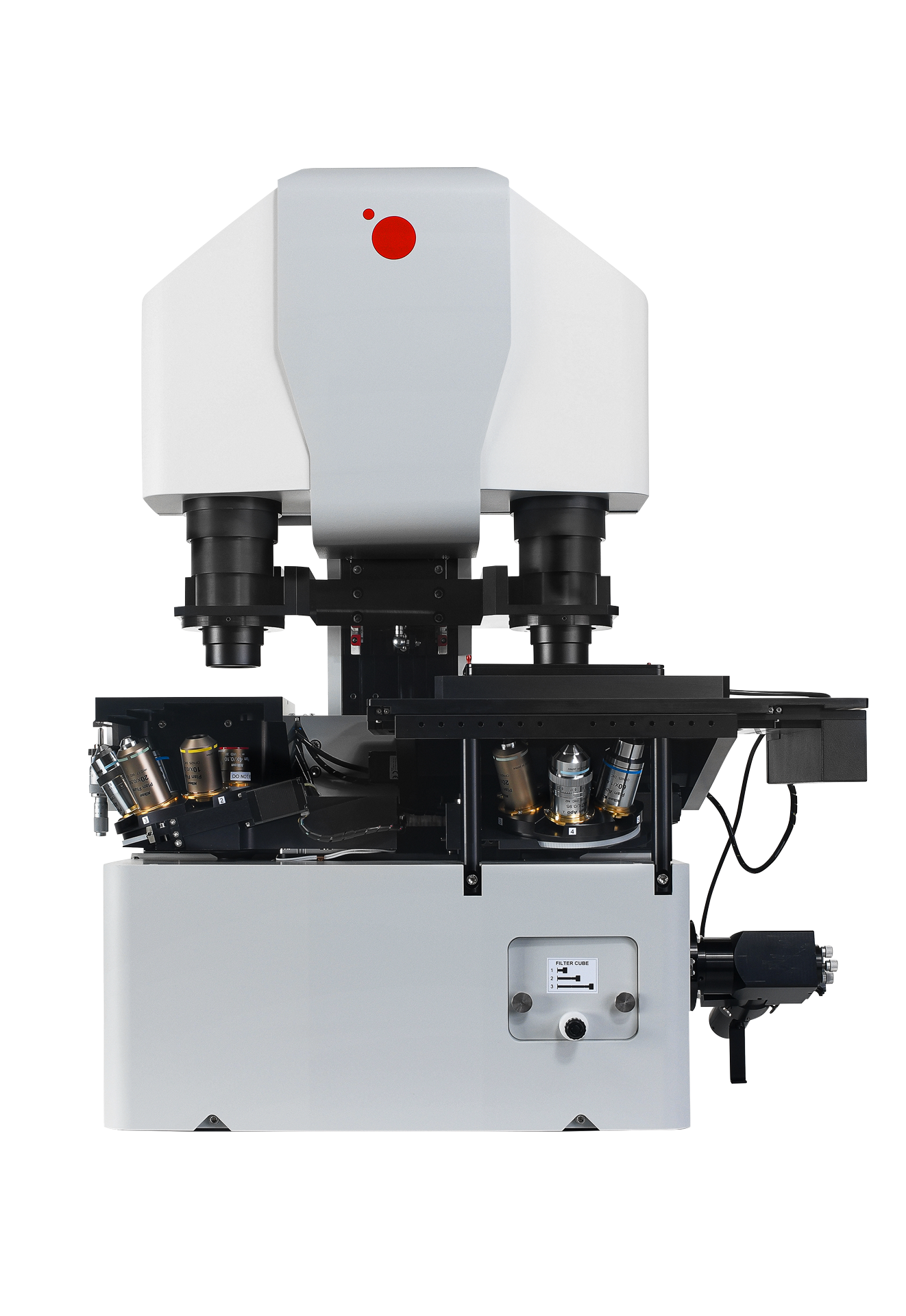
Q-Phase
Quantitative Phase Imaging
Q-Phase is a patented holographic microscope with high detection sensitivity, designed for gentle live-cell imaging.
Q-Phase is an ideal solution for experts who desire precise automated segmentation of individual cells for subsequent data analysis. Q-Phase quickly transforms cell features and dynamics into numerical data suitable for comparisons, correlations, and more detailed statistics.
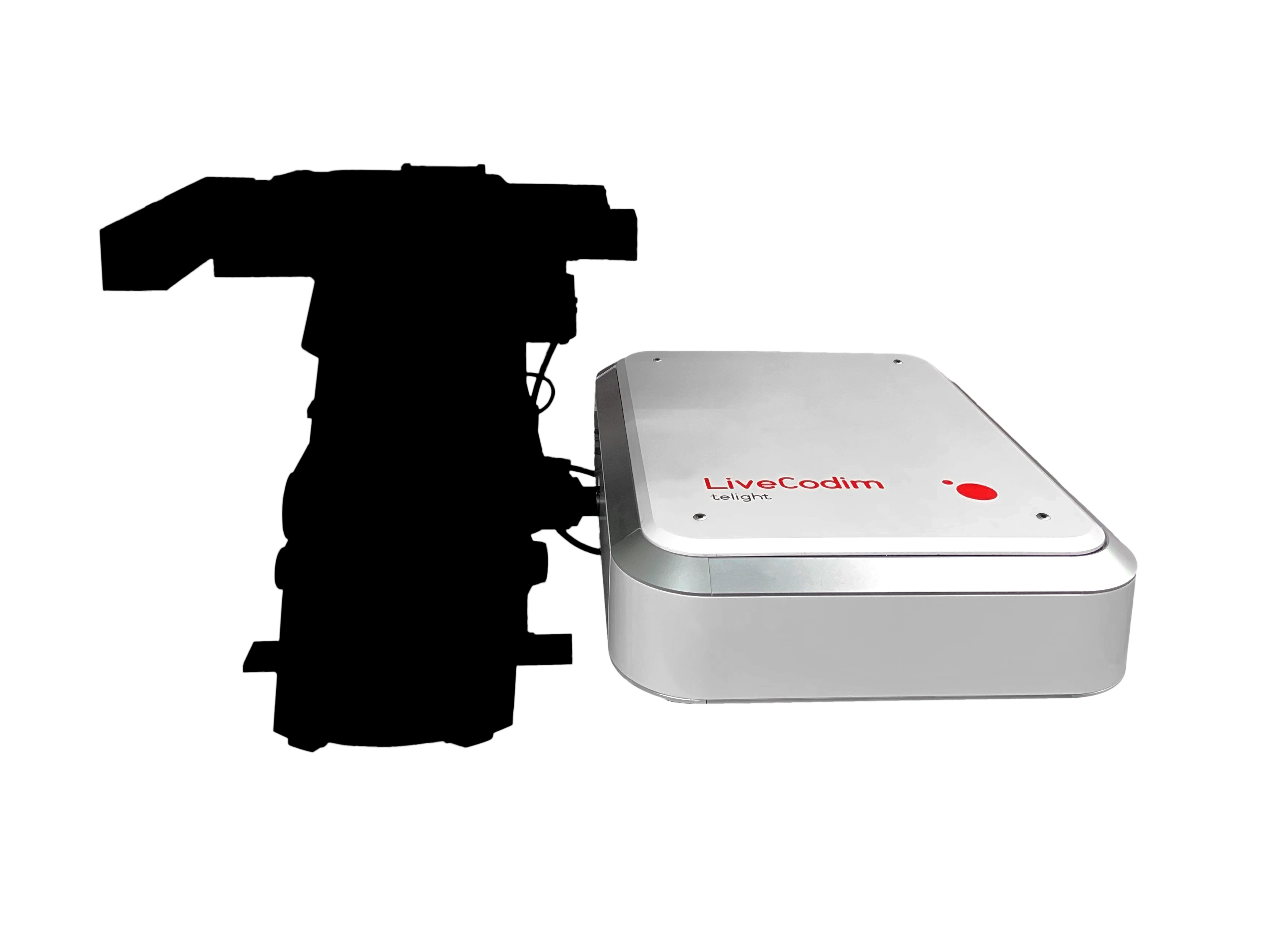
LiveCodim
From conventional to super-resolution microscopy
LiveCodim is a universal, super-resolution imaging platform designed to interface with any standard fluorescence microscope. It is the solution for live-cell imaging with high resolution and low phototoxicity.