

Immunology
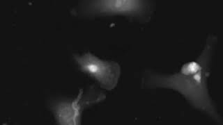
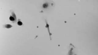
Dysregulation of immune cell functions can result in many serious and fatal diseases that require re-modulating the immune cells and restoring their normal response. Monitoring of immune cells while interacting with self-cells, pathogens, and cancer promotes our understanding of immune response or its failure mechanisms.
In recent years, immunotherapy gained high potential in cancer treatment as engineered T cells represent a powerful new approach for targeting cancer and autoimmune disorders. However, there are concerns about safety and off-target toxicity, as well as the development of resistance.
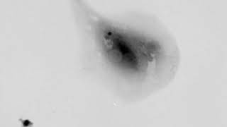
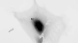
Q-Phase opens new horizons for studying spatiotemporal mechanisms and interactions of immune cells under label-free conditions, thus enabling various applications in research.

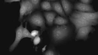
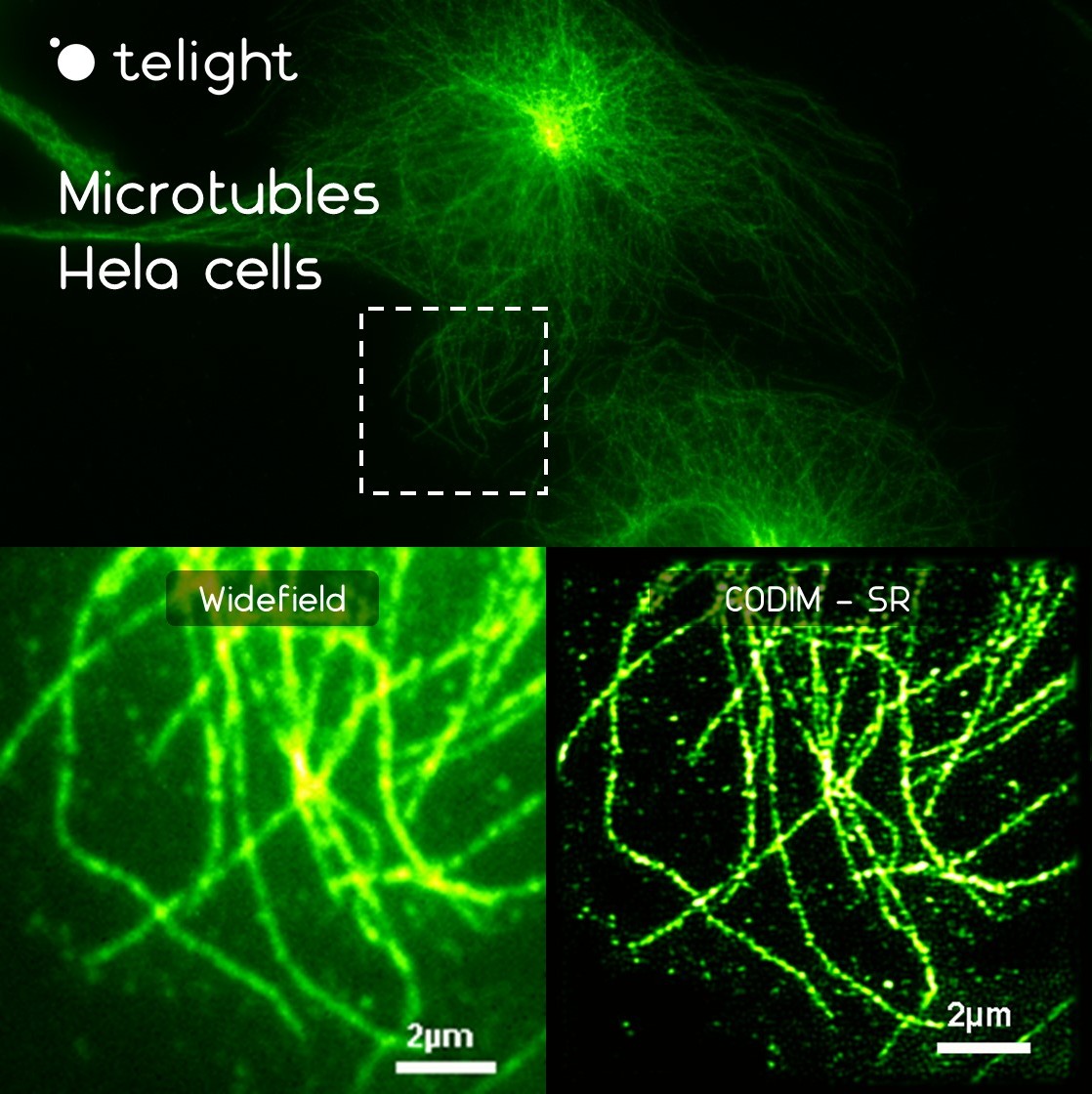
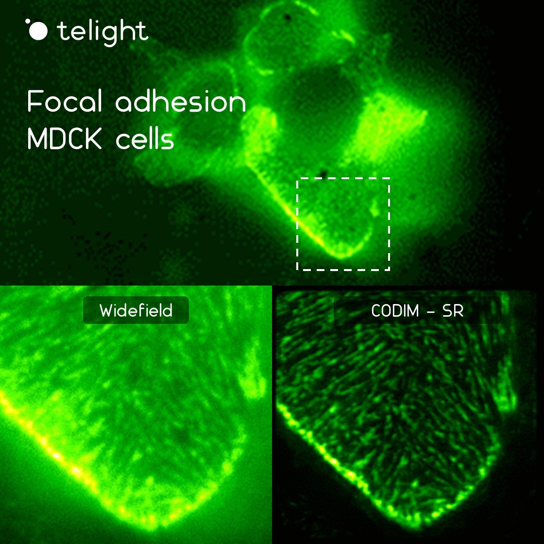
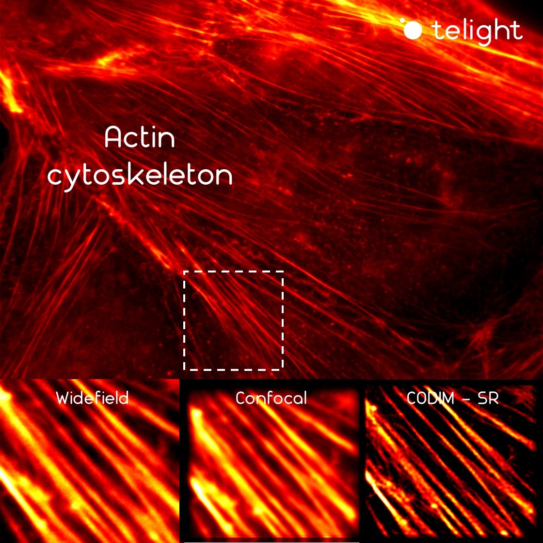
Super-resolution microscopy is crucial for investigating cell-cell interaction and particularly, microbiological immunity (against bacteria and viruses).
Indeed, due to the small size (few nm to µm) of microbes, studying immune responses on cellular and molecular levels remains challenging in conventional optical microscopy, but this drawback could be overcome using the LiveCodim super-resolution module.
Publications
Maarifi, G., Fernandez, J., Portilho, D.M., et al.
RanBP2 regulates the anti-retroviral activity of TRIM5α by SUMOylation at a predicted phosphorylated SUMOylation motif
Products
Telight Q-Phase
Quantitative Phase Imaging
Q-Phase is a patented holographic microscope with high detection sensitivity, designed for gentle live-cell imaging.
Q-Phase is an ideal solution for experts who desire precise automated segmentation of individual cells for subsequent data analysis. Q-Phase quickly transforms cell features and dynamics into numerical data suitable for comparisons, correlations, and more detailed statistics.
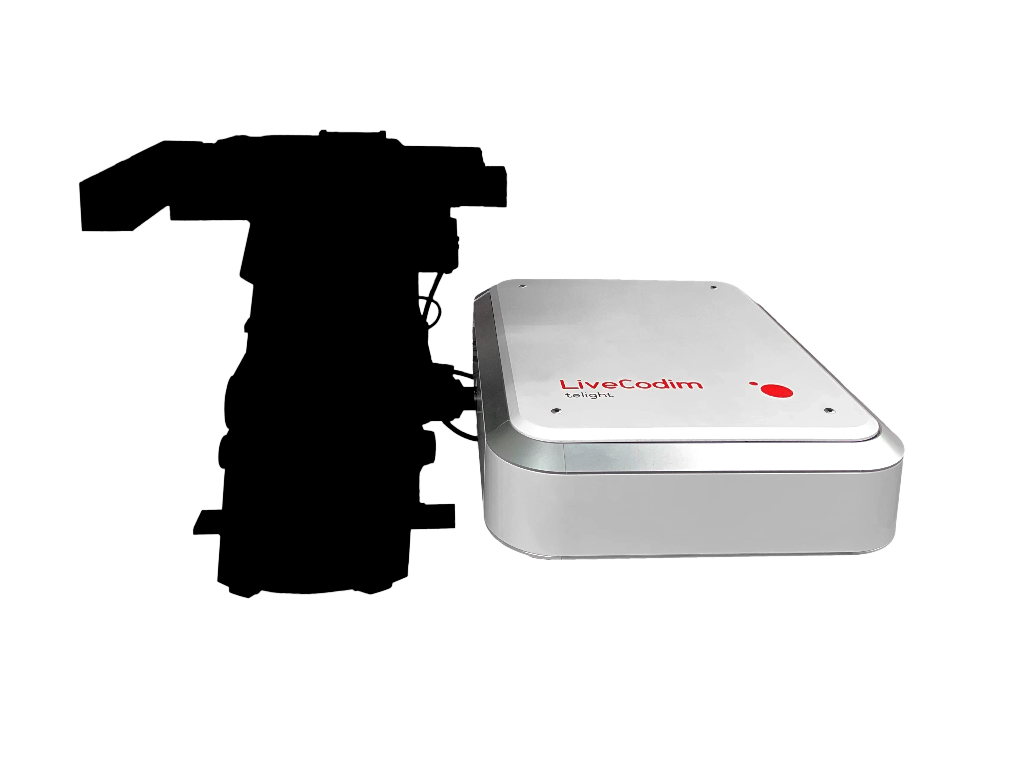
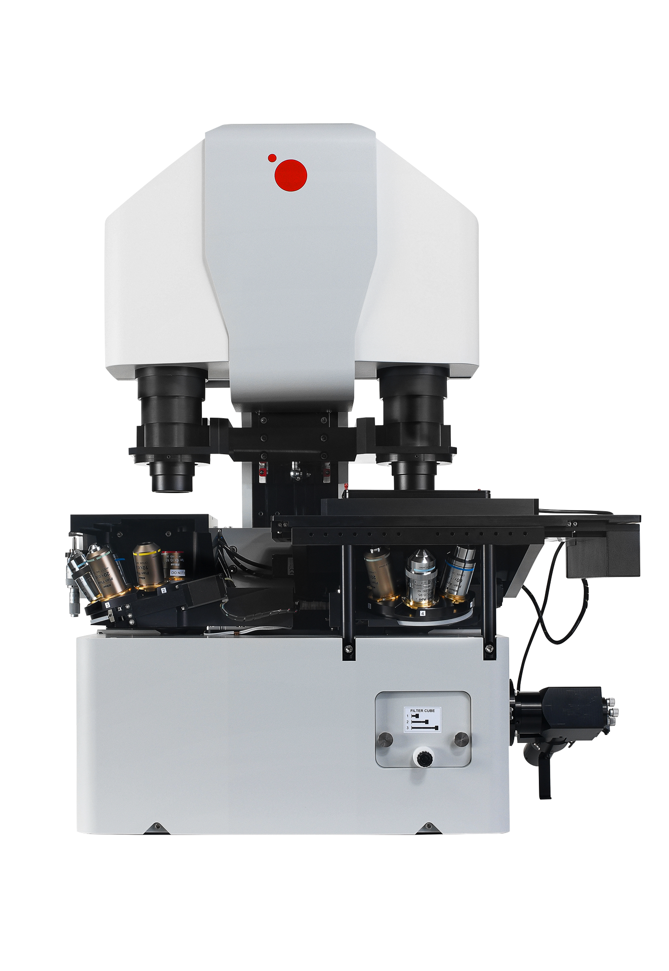
Telight LiveCodim
From conventional to super-resolution microscopy
LiveCodim is a universal, super-resolution imaging platform designed to interface with any standard fluorescence microscope. It is the solution for live-cell imaging with high resolution and low phototoxicity.