

Cancer research
Cancer is an incredibly diverse disease that can arise in different organs and tissues. This diversity is not limited to different tumor types but cells within the same tumor can also vary.
Tumor heterogeneity represents a major medical challenge that requires innovative methods for understanding the role of each distinctive cell population in promoting tumor growth, resistance, and progression.
Q-Phase
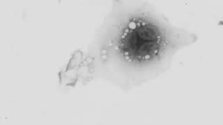
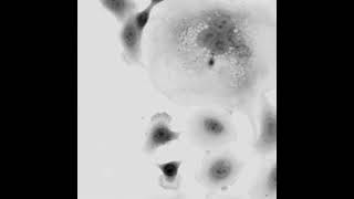
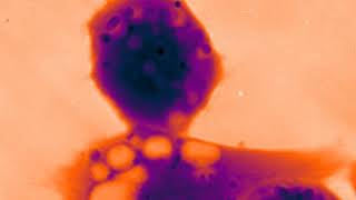
A comprehensive tool to investigate individual cells within a tumor is essential. Q-Phase provides a new opportunity that enables researchers to interpret multiple cellular parameters in real-time including cell morphology profiling, cell dynamics, and cell dry mass.
Live-cell imaging using Q-Phase allows for automated and precise segmentation of cells outlines with subsequent credible statistics and correlations. Therefore, assessment of cell cycles, cell death, and cell migration can be closely achieved in a single experiment.

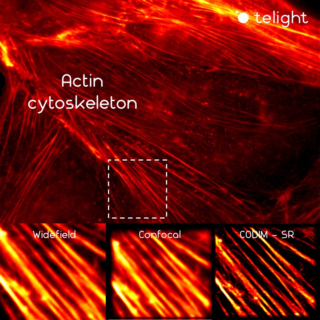
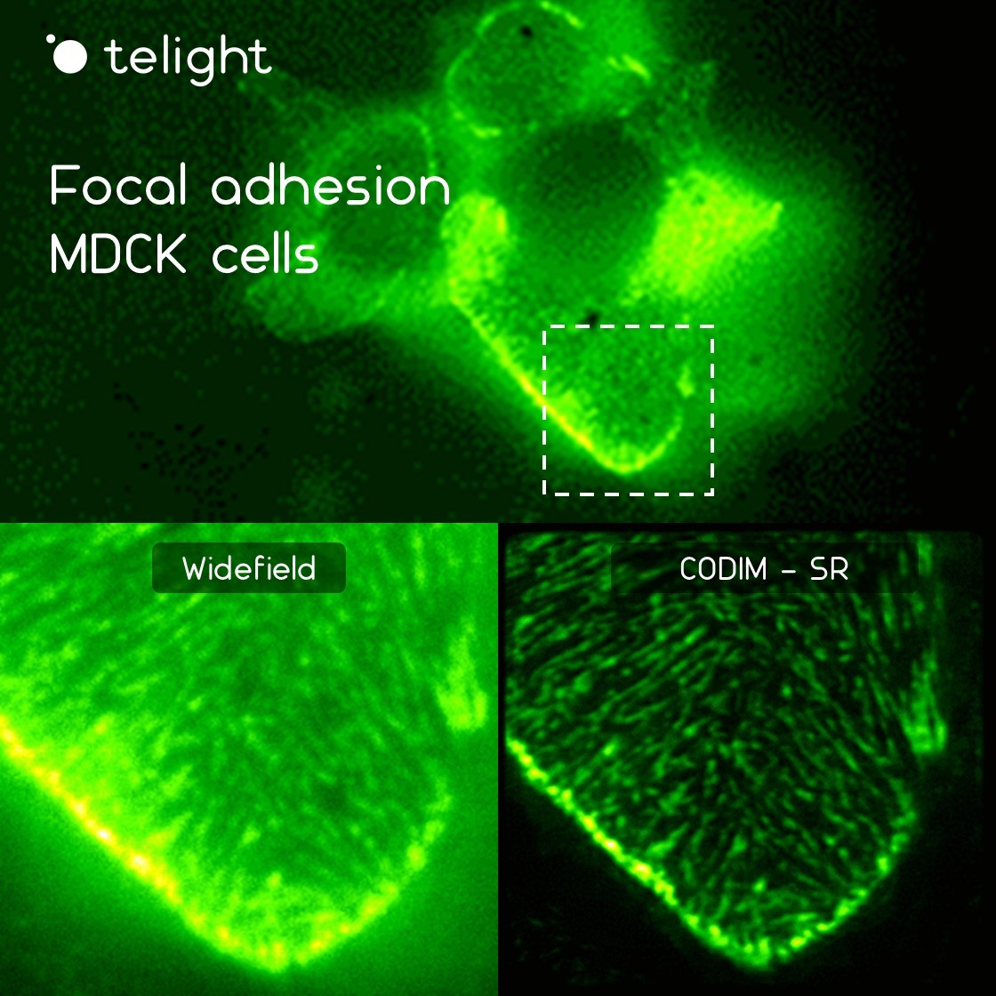
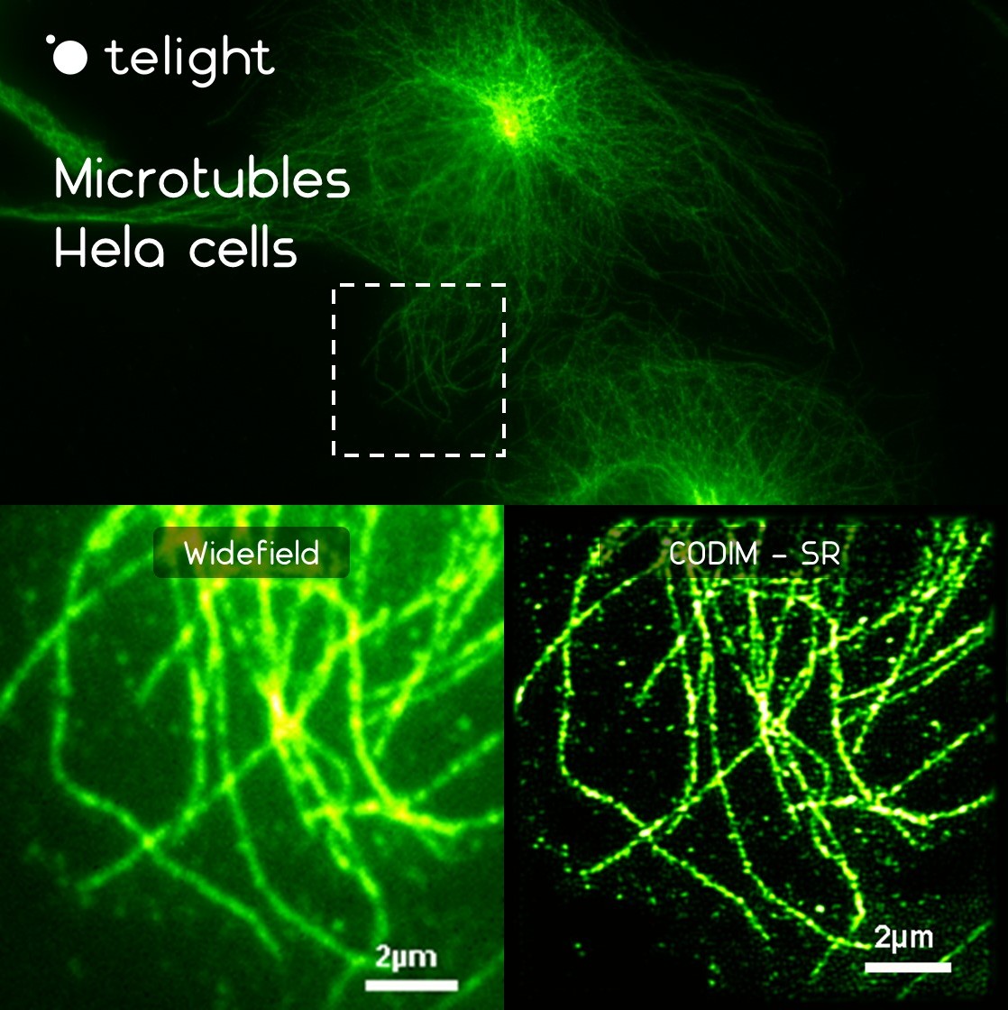
LiveCodim
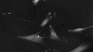
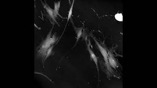
During the last decades, microscopy became a weapon of choice to elucidate cancer-cell biology processes such as the steps and development of metastasis.
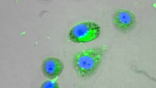
The LiveCodim module is of interest in this context as it provides new visualization of living cancer cells in high resolution. For example, LiveCodim can bring new insights into the visualization of mitochondria, cytoskeleton, nuclei dynamic, vesicle trafficking, etc.
Publications
M. Fojtů, et al.
Silicane Derivative Increases Doxorubicin Efficacy in an Ovarian Carcinoma Mouse Model: Fighting Drug Resistance
Quantitative Phase Dynamics of Cancer Cell Populations Affected by Blue Light
J. Balvan, et al.
Oxidative Stress Resistance in Metastatic Prostate Cancer: Renewal by Self-Eating
Cisplatin enhances cell stiffness and decreases invasiveness rate in prostate cancer cells by actin accumulation
Products
Q-Phase
Quantitative Phase Imaging
Q-Phase is a patented holographic microscope with high detection sensitivity, designed for gentle live-cell imaging.
Q-Phase is an ideal solution for experts who desire precise automated segmentation of individual cells for subsequent data analysis. Q-Phase quickly transforms cell features and dynamics into numerical data suitable for comparisons, correlations, and more detailed statistics.
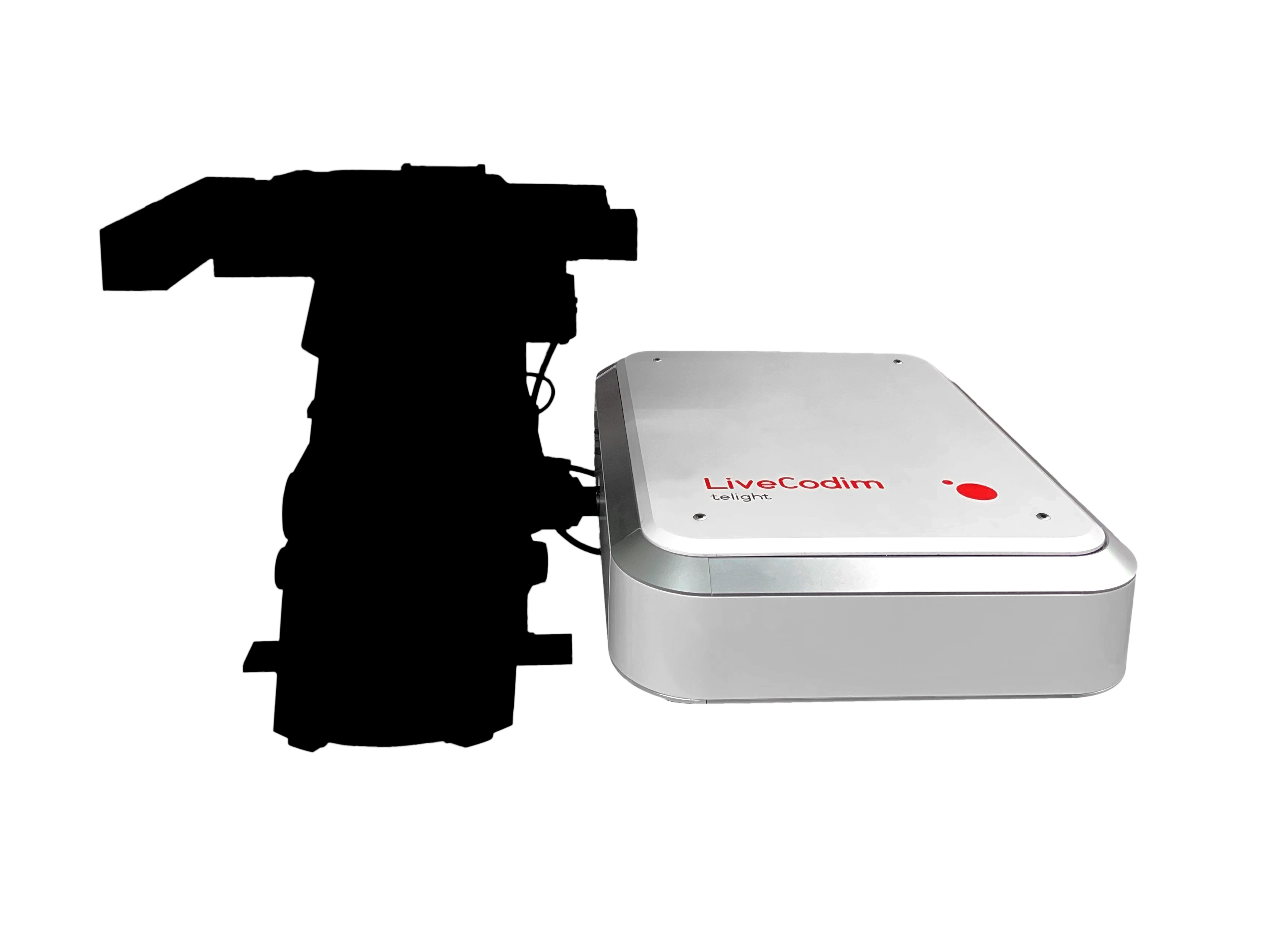
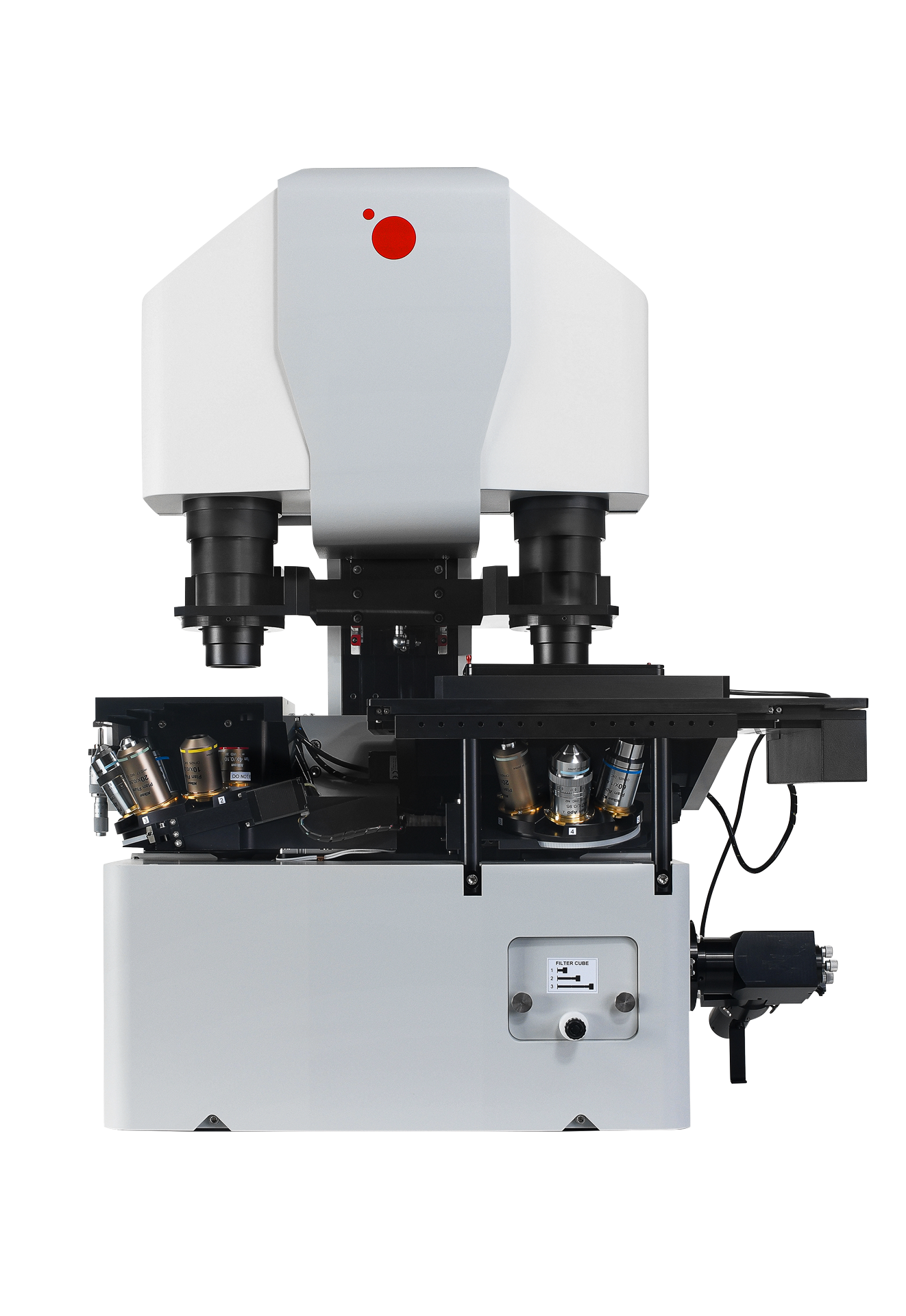
LiveCodim
From conventional to super-resolution microscopy
LiveCodim is a universal, super-resolution imaging platform designed to interface with any standard fluorescence microscope. It is the solution for live-cell imaging with high resolution and low phototoxicity.