Biological research is constantly evolving, driven by new technologies that expand our ability to observe and understand living systems. At Telight, we develop advanced instruments for this purpose, including the Q-Phase holographic microscope, a tool designed to capture the dynamics of life at the cellular level with exceptional precision. As research increasingly depends on data analysis, we are integrating artificial intelligence and machine learning into our quantitative phase imaging (QPI) software SophiQ.
The power of QPI and AI in modern life sciences
Quantitative phase imaging provides label-free and non-invasive imaging of living cells while directly measuring their dry mass, a fundamental parameter linked to cell physiology, metabolism, and disease progression. The use of incoherent light in the Q-Phase microscope ensures artifact-free, uniform data, creating ideal conditions for analysis with artificial intelligence. This consistency enables the use of deep learning algorithms for automated detection, segmentation, tracking, and classification of cells and intracellular structures.
When combined with artificial intelligence, quantitative phase imaging becomes a powerful tool for phenotypic analysis, drug screening and disease modeling. Machine learning models trained on large datasets can detect subtle morphological or biophysical changes that remain invisible to the human eye, revealing early cytotoxic effects in drug testing or slight variations in cell growth and migration associated with cancer research.
Transforming research with data-driven insight
The integration of artificial intelligence into quantitative phase imaging data processing shows great promise for a wide range of research applications, including cancer research, immunology and drug discovery. Automated algorithms can recognize and quantify cell behavior, improving for example the accuracy of toxicity assessments, screening of new compounds, and studies of immune cell interactions. This approach not only accelerates experiments but also enhances reproducibility and objectivity in biomedical research.
Advanced image analysis with a machine learning approach
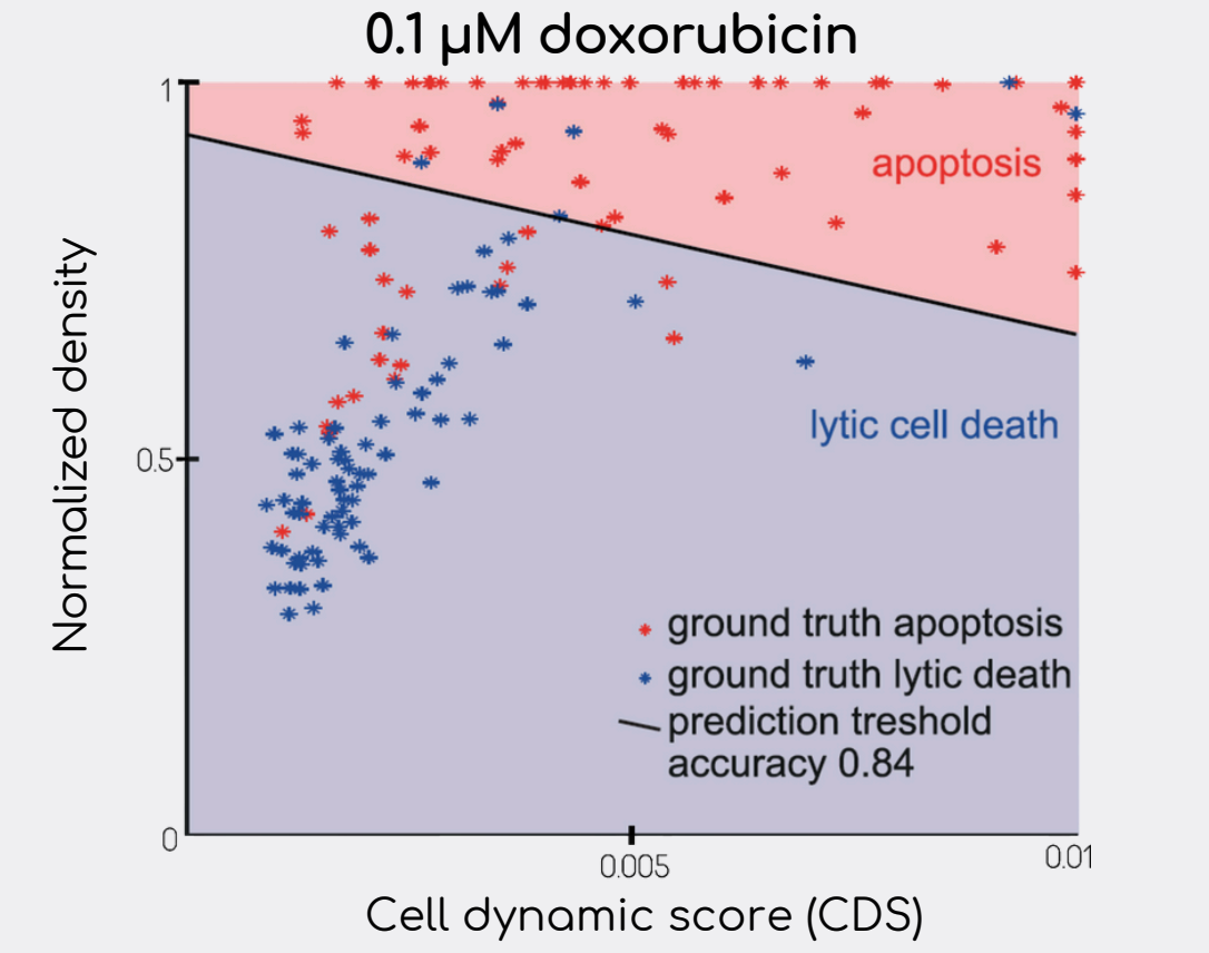
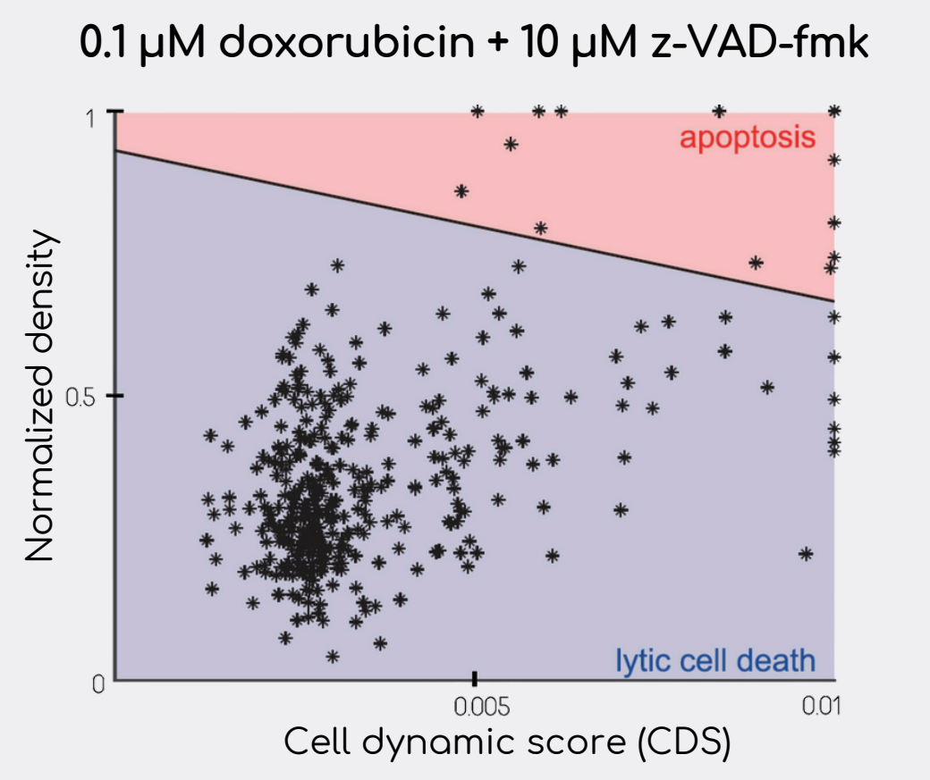
This study highlights an example of how AI models trained on QPI data can automatically detect and classify cell death [2]. Scientists applied the long short-term memory (LSTM) neural network to automatically detect the time point of cell death from extracted features (mass, area, density, etc.) of segmented and tracked LNCaP, DU-145, and PNT1A cells. For cell death type classification, only two features were selected: mass density (pg/pixel) and cell dynamic score (CDS), features introduced by the authors. These features served as an input for the machine learning algorithm for cell death distinction. As a result, their method identified cell death subroutines based on patterns in the time-lapse QPI data, achieving 75.6 % accuracy for DU-145 cells compared to a manual detection method depending on fluorescence signal analysis.

Classification of cell death type in DU-145 cells (N = 160) based on the automatic detection method. Blue points represent manually annotated necrotic cells. Red points are manually annotated apoptotic cells. Cells were treated with doxorubicin. Original graph from [2].

Classification of DU-145 cells based on an automatic detection method without manual annotation. Each point represents one cell (N = 381). Inhibitor of apoptosis z-VAD-fmk causes increased progression of lytic cell death (necrosis). Original graph from [2].
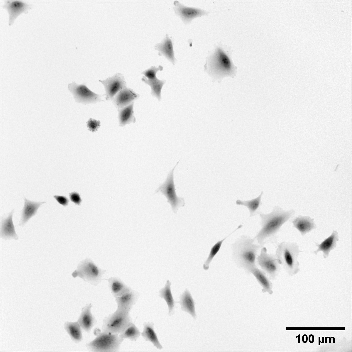
Deep learning further allows advanced segmentation and feature extraction, enabling researchers to study cell morphology, dynamics, and structure with remarkable precision. Artificial intelligence can label organelles or predict cellular changes over time without fluorescence staining, reducing phototoxicity and preserving natural cell function during long-term experiments.
Example of the segmentation mask used in the model training for SophiQ:


Collaboration with the European Digital Innovation Hub in Ostrava
From data to discovery
By combining the quantitative strength of QPI with the adaptive intelligence of machine learning, Telight provides researchers with a tool that can segment vast amounts of biological data efficiently and reliably. This synergy leads to faster discoveries in fields such as cancer research, drug toxicity testing, migration studies, or plant cell biology, and contributes to a more comprehensive understanding of life itself.
At Telight, we continue to refine these capabilities to make artificial intelligence-enhanced quantitative phase imaging not only a method of observation but also a platform for quantitative, data-driven discovery in the life sciences.
QPI data and your research
If you are interested in the data that were produced by our Q-Phase microscope, we have multiple datasets available to share with you, so you can see the quantitative data and run your models on them. Our application specialists are always glad to talk about your research. Do not hesitate and contact us for demo or showcase of our software capabilities. You can download our full application note on AI and Quantitative Phase Imaging.
References
[1] Vicar, T. et al. Cell segmentation methods for label-free contrast microscopy: review and comprehensive comparison. BMC Bioinformatics 20, 360 (2019).
[2] Vicar, T., Raudenska, M., Gumulec, J. & Balvan, J. The Quantitative-Phase Dynamics of Apoptosis and Lytic Cell Death. Sci. Rep. 10, 1566 (2020).