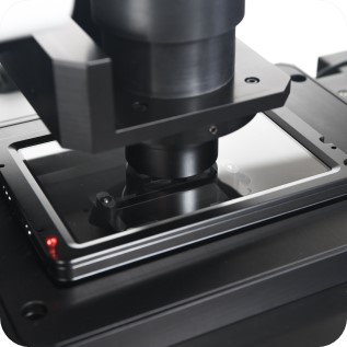
Telight Q-Phase
Quantitative Phase Imaging
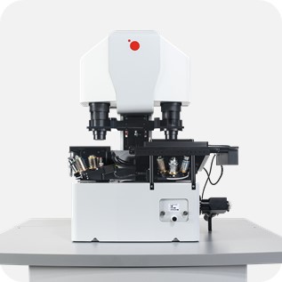
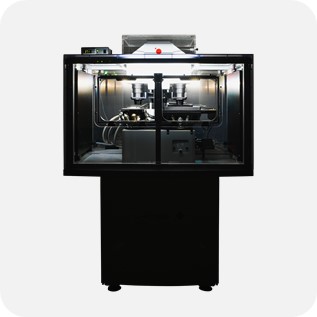
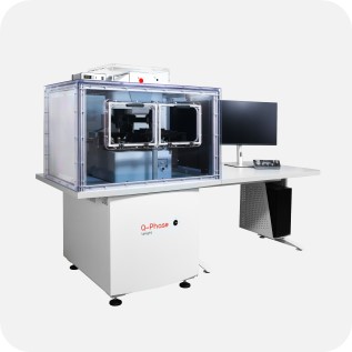
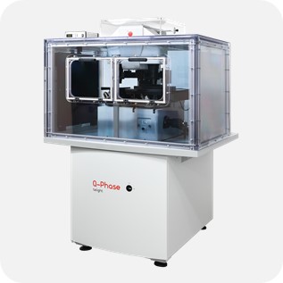
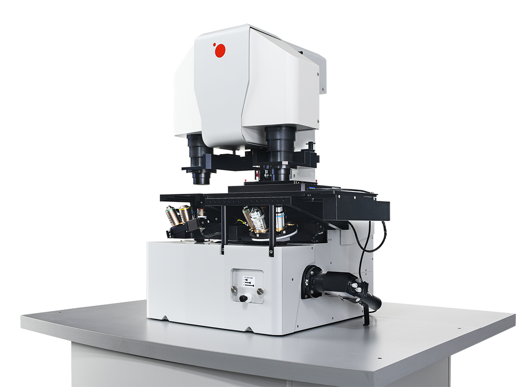
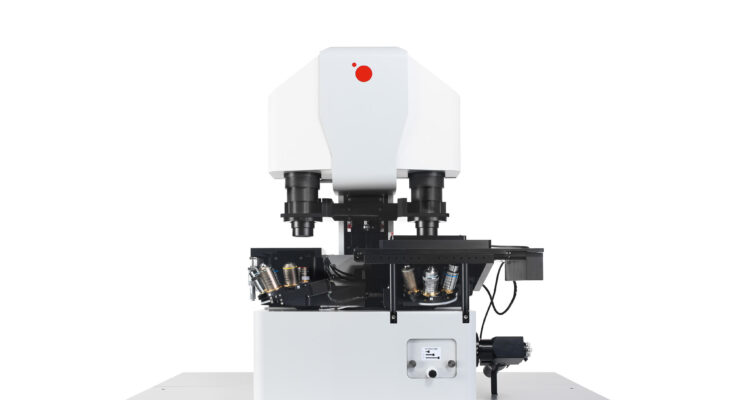
Q-Phase is a patented holographic microscope with high detection sensitivity, designed for gentle live-cell imaging.
Q-Phase is an ideal solution for experts who desire precise automated segmentation of individual cells for subsequent data analysis. Q-Phase quickly transforms cell features and dynamics into numerical data suitable for comparisons, correlations, and more detailed statistics.
Description
Q-Phase: Cell culture analysis
Q-Phase is a quantitative label-free live-cell imaging system - ideal if you desire reliable automated segmentation and analysis of cell behavior. It is a holographic microscope optimized for real-time monitoring of living cells with minimal phototoxicity. Q-Phase easily transforms cell features and dynamics into reliable data for your analysis and correlations.
All-steps-in-one
Q-Phase was purposely designed to observe living cells in vitro for long-term several-day experiments. The system creates space for observation, automated, precise, and fast cell segmentation, and thanks to software specifically developed for Q-Phase also analysis – all steps in one system.
Observe
Q-Phase solution is based on the unique patented technology of Coherence-Controlled Holographic Microscopy (CCHC). This technology uses incoherent light sources (halogen lamp or LED) providing QPI with high quality. Moreover, it is the only QPI technique, that enables imaging of samples in scattering media (phospholipid emulsions, extracellular matrices, etc.)
- Unique cellular contrast – The quantitative phase imaging directly visualizes cellular mass (thickness x refractive index).
- No image artifacts such as halo effect (as opposed to techniques based on Zernike phase contrast illumination).
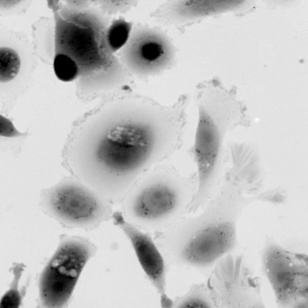
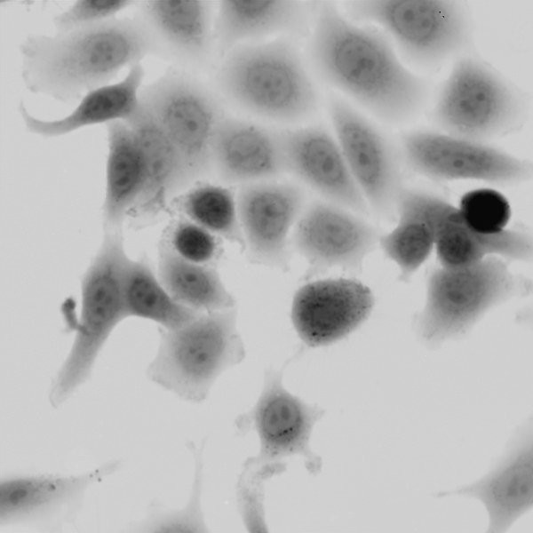
- Transparent cells made visible – Even the most transparent cells and their outlines can be distinguished from the background.
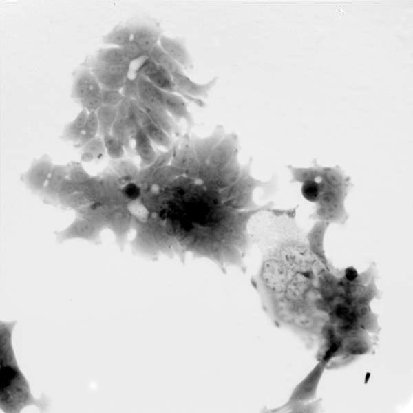
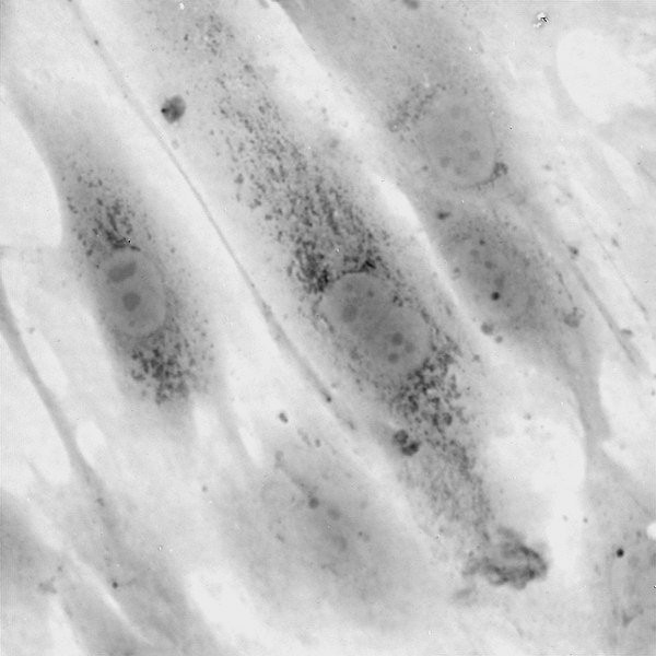
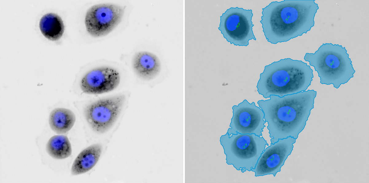
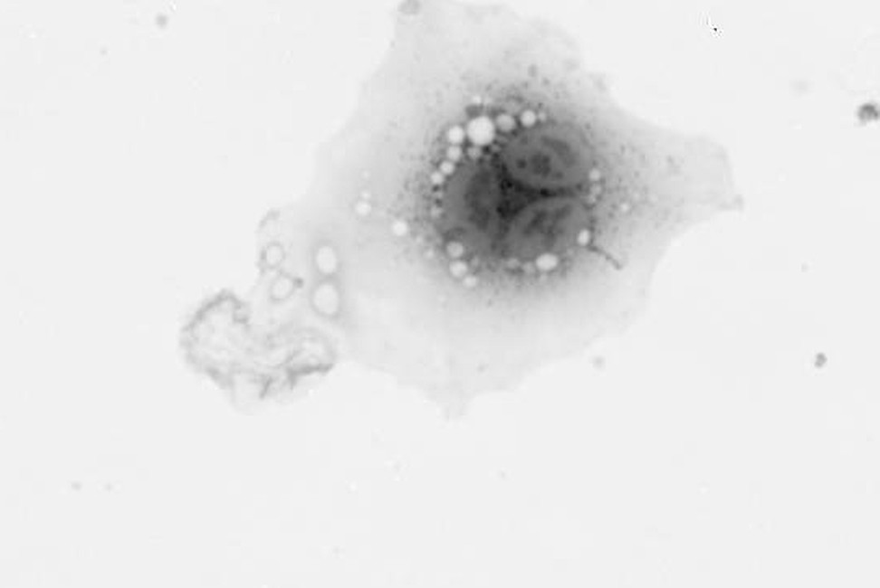
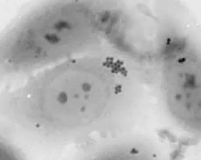
- Look inside the cells – Changes in internal parts of cells such as nuclei vacuoles, and many more, can be seen without any labeling.
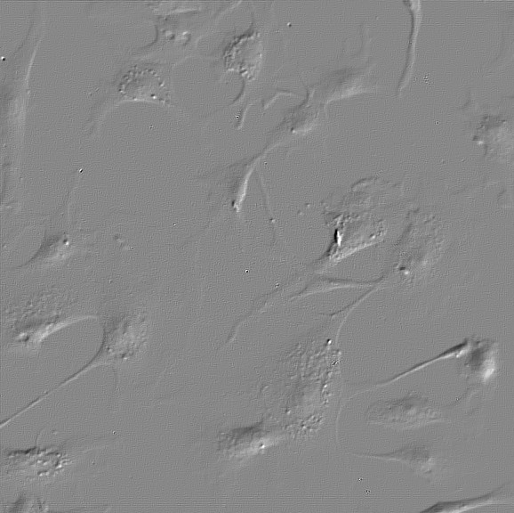
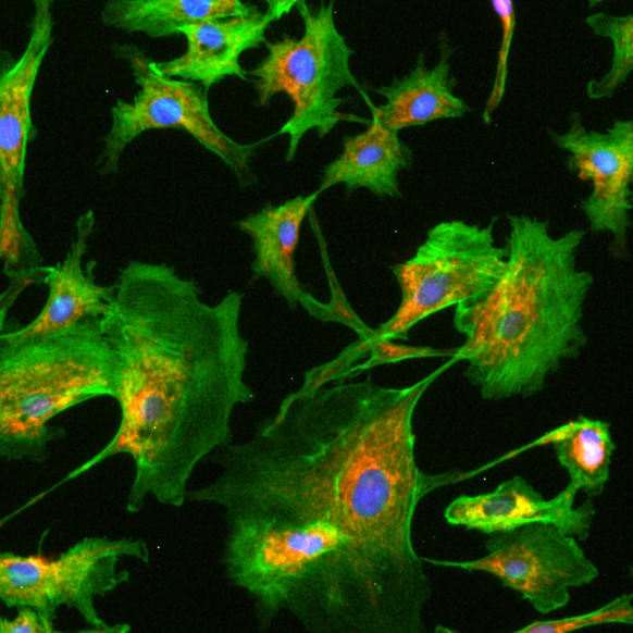
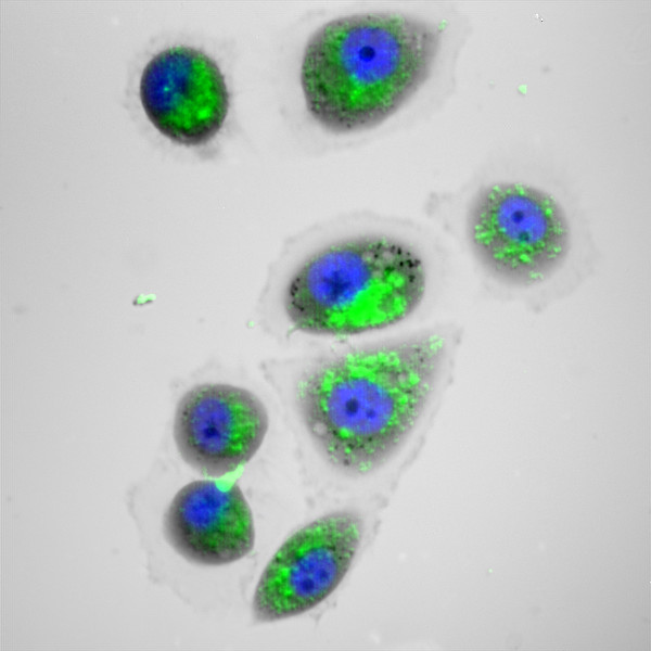
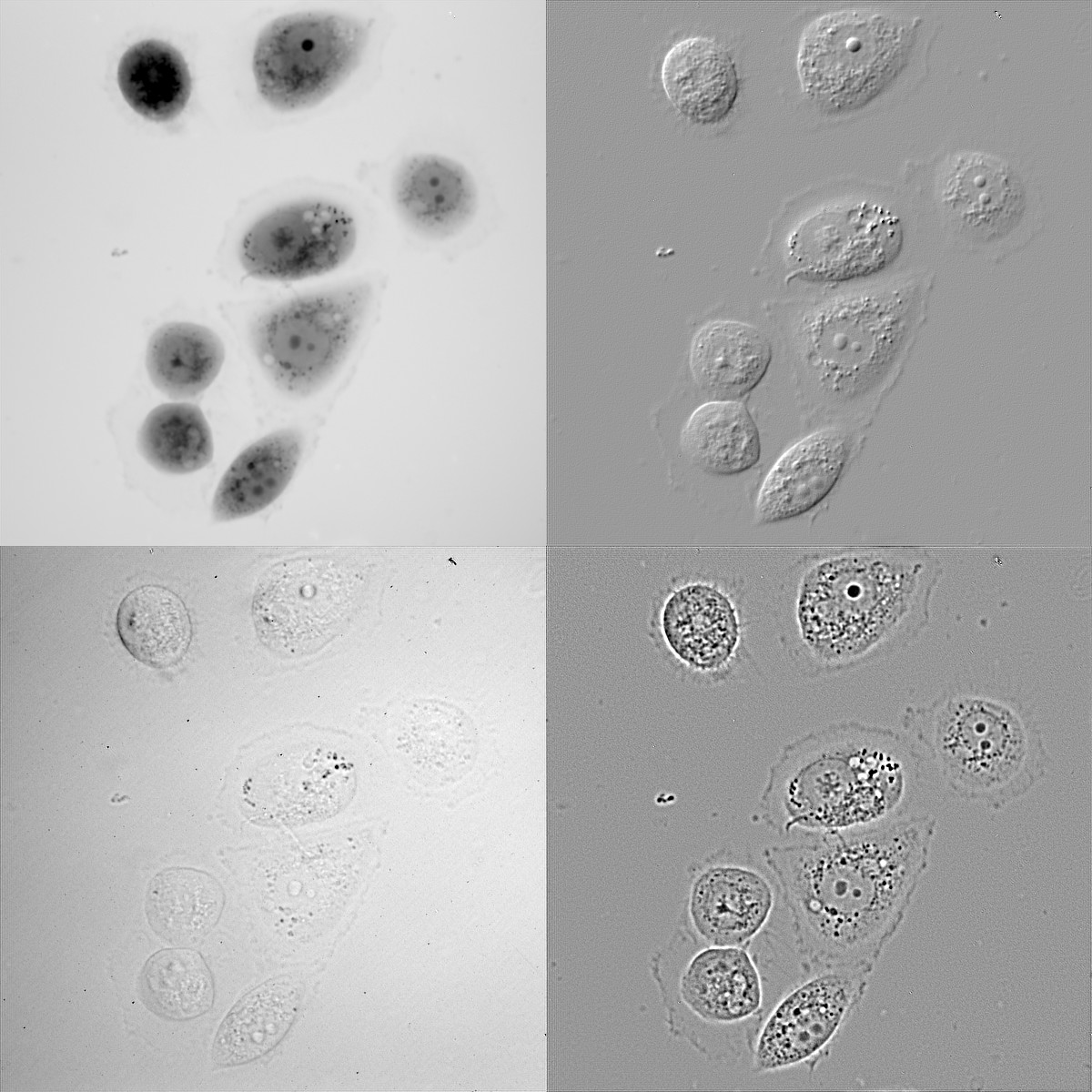
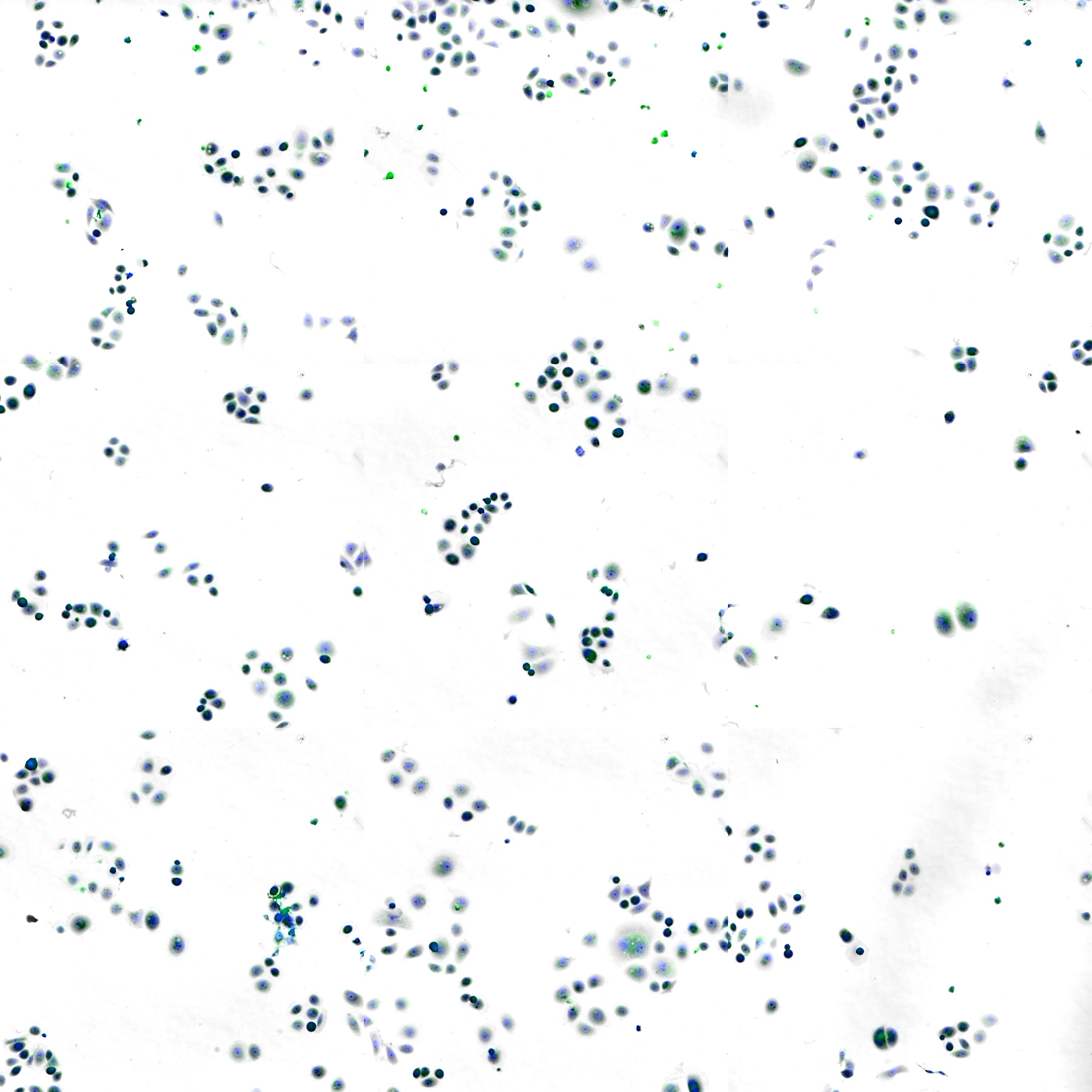
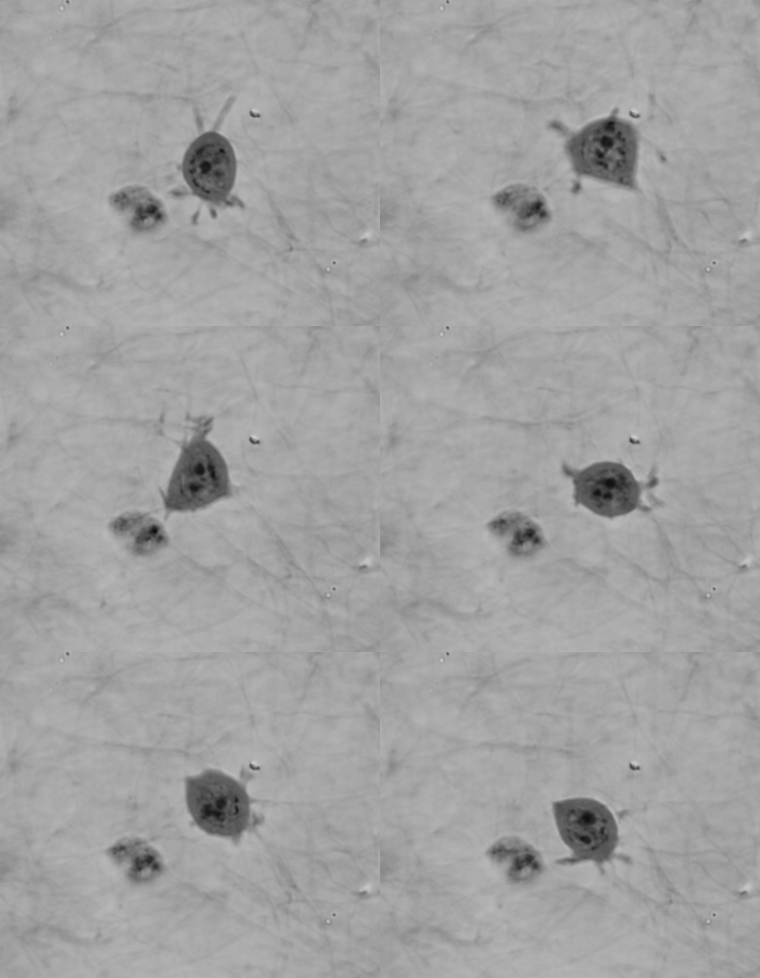
- Multiple imaging modes – Additional imaging modalities are available such as widefield fluorescence, simulated DIC, brightfield, or high-pass filtered QPI. Multiple dimensions can be combined in a single experiment and automatically acquired by the Q-Phase system (timelapse, multi-position, multi-channel, Z-stack) to provide a more complete picture in a synergistic fashion.
Automatically SEGMENT the data with high accuracy
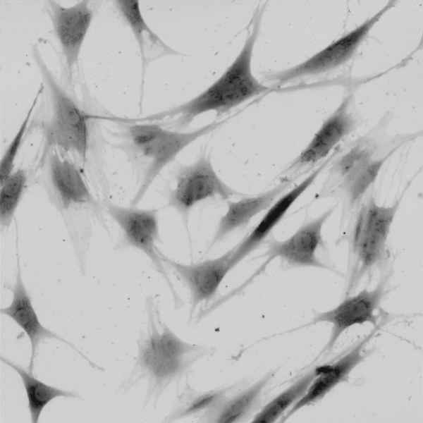
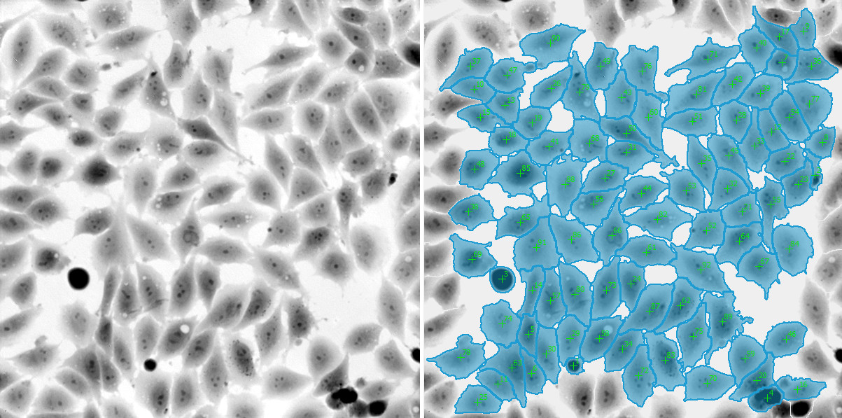
- QPI images offer the highest cell/background contrast among other label-free imaging techniques thus enabling the most reliable and precise automated (or manual) segmentation of individual cells. Even highly confluent populations can be reliably segmented.
- Moreover, fluorescence data can be utilized to refine the segmentation even further.
- QPI-based segmentation is also very fast, which significantly speeds up analyses of large datasets with thousands of frames.
ANALYZE quantitative data for every cell
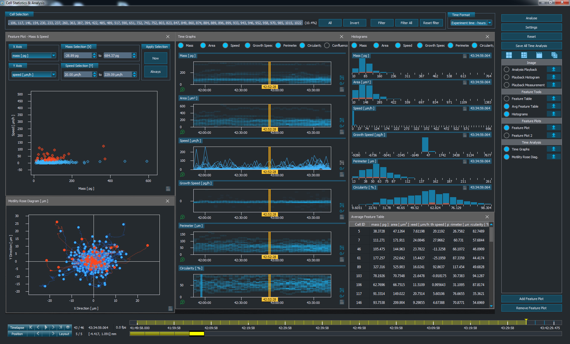
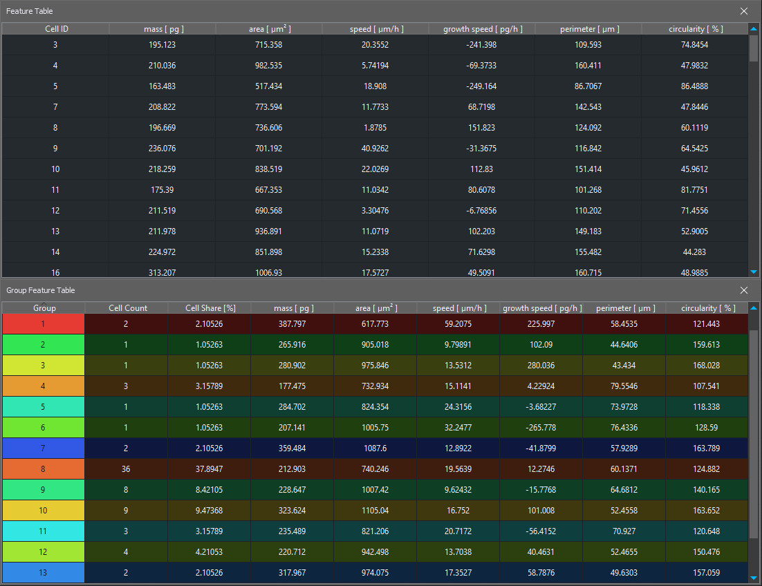
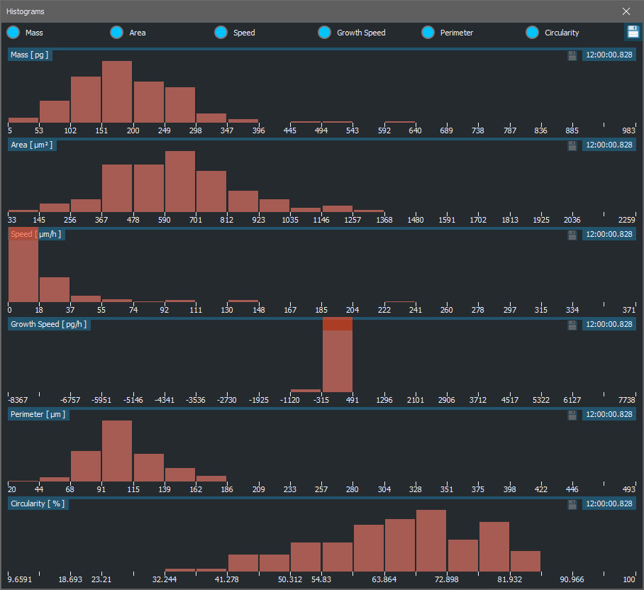
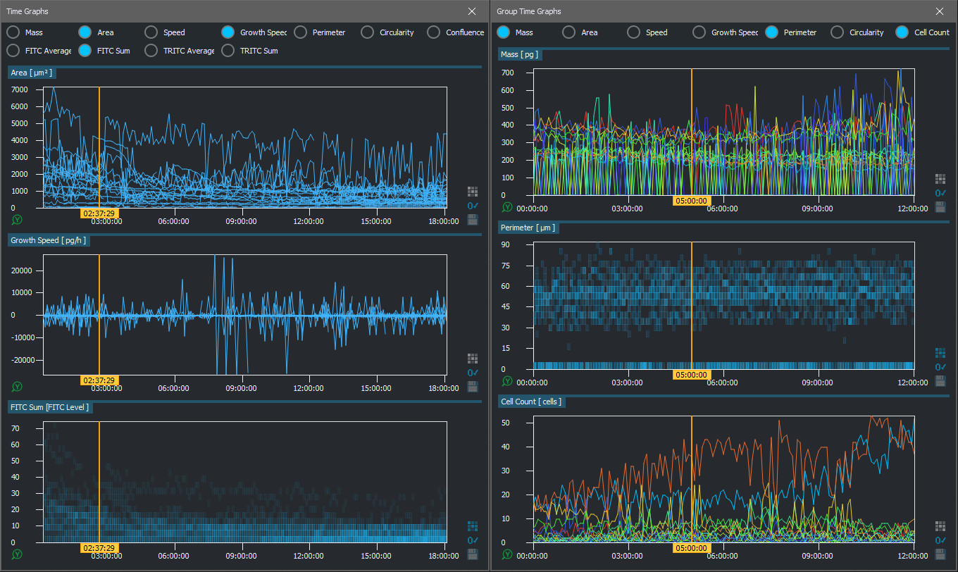
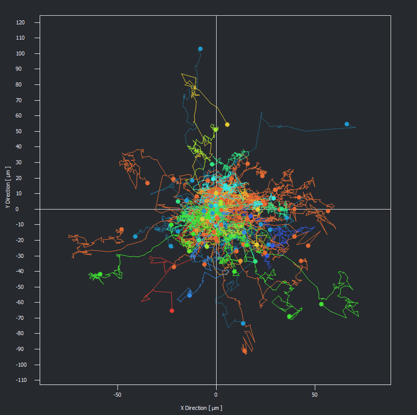
- The powerful Q-Phase analysis toolbox processes segmented images on-the-fly and provides a complex portfolio of tools for data visualization, subpopulation gating, and multi-parameter data mining. It links quantitative data to images and individual cells, which makes optimizing gates and checking outliers extremely easy and efficient.
- The resulting data can be exported in common file formats for further processing and analysis.
Key advantages
AUTOMATED SEGMENTATION, precise detection of cell boundaries – reliable and fast, comparable to fluorescence data processing
Phase values can be recalculated e.g. to CELL DRY-MASS DENSITY (pg/μm2) or direct topography with nanometer sensitivity (typically non-biological samples with homogeneous refractive index distribution)
FULLY INTEGRATED solution | All-steps-in-one
SophiQ - software strongly supporting analysis | Providing all necessary steps
Technology feasible for cell monitoring IN SCATTERING MEDIA
QPI + Fluorescence Imaging = Experiments monitoring cells FOR WEEKS
Download
Publications
S. Lewis, et al.
A mechanism for MEX-5-driven disassembly of PGL-3/RNA condensates in vitro
Velluvakandy, et al.
Nanorobot-Cell Communication via In Situ Generation of Biochemical Signals: Toward Regenerative Therapies
Chvalová, et al.
Comparison of holotomographic microscopy and coherence-controlled holographic microscopy
Suranova, et al.
Primary assessment of medicines for expected migrastatic potential with holographic incoherent quantitative phase imaging
Technology and software
Quantitative Phase Imaging (QPI) is a novel label-free imaging technique bringing a completely new contrast into the gentle live cell imaging field.
Q-Phase imaging allows the extraction of information-rich quantitative data from unlabelled cells. It is directly proportional to the cellular dry mass and can be used for monitoring and direct quantification of dry mass changes inside the cells in pg/μm2. Even the slightest mass changes can be detected and quantified thanks to the high sensitivity.
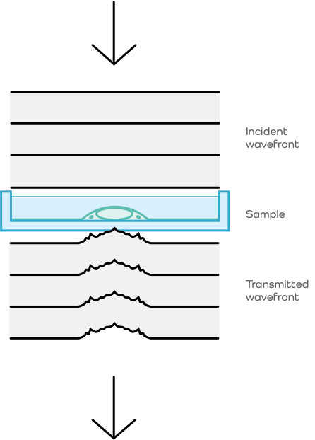
Principles of quantitative phase imaging
The time of propagation of light in a specific environment depends on the refractive index as well as the distance of the optical path. Therefore, when a light wave travels through a sample with varying refractive index and/or height, its wavefront is distorted and a change in the phase distribution of this wave occurs. The Q-Phase allows detecting the phase distribution in the sample plane. This process of phase-detection at a sample plane is usually referred to as quantitative phase imaging.
Quantitative phase image can provide information on sample morphology, topography, or cell dry-mass distribution. Cell dry mass is quantified in pg/μm2 and can be calculated directly from phase values detected in each pixel. Quantitative phase imaging provides a very simple and sensitive way for monitoring cell reactions to treatment and analyses of growth, area, shape, movement, and many other parameters.
Patented optical setup
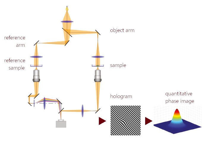
The Q-Phase microscope consists of two arms, an object arm, and a reference arm. The arms have similar microscope setups with a common illumination system. The sample is placed into the object arm, and the so-called reference sample (blank) is placed into the reference arm. The beams in each arm pass through the inserted sample and are combined at the image plane of the microscope.
Thanks to the Q-Phase´s unique patented optical setup, the beams interfere and form a hologram even when illuminated with a halogen lamp or a LED. The hologram is then recorded by a detector and a quantitative phase image is extracted from the hologram in real-time by a computer.
The origins of Q-Phase
The invention of Q-Phase is attributed to Prof. Radim Chmelík, who is currently working at CEITEC as a Research Group Leader at the Experimental Biophotonics Group. In the 90s, together with researchers from Brno Technical University, he came up with a revolutionary thought of constructing a confocal microscope without scanning based on incoherent-light-source holographic microscopy. This was the basis for the optical set-up of the Q-Phase microscope. The patented technology has brought him and his colleagues several awards, such as the Werner von Siemens Excellence Award in 2013 and ‘Česká Hlava’ (Czech Brains), Kapsch Invention Prize category in 2016.
We place a strong emphasis on collaboration with academia, which is why we continue to develop our instruments in close partnership with leading Czech technical universities, scientists, and research institutions.
SophiQ - 2 modes of software support for your easy work
Live mode
- Complex experiment design (automated acquisition of up to 4 dimensions – time, position, channel, Z-stack)
- User management – personalizable setup of analysis, user history tracking
Dataset mode – offline view and work with images
- Comprehensive & interactive data analysis toolbox (automated cell segmentation, scatter plots with population gating, time plots, histograms, data tables etc.)
- Multidimensional data viewer
- GPU accelerated image processing
- Direct export to video
Plus
ImageJ plugin – open Q-Phase datasets directly in ImageJ
Automatic updates that enable you to keep up with the newest features
Free viewer lets you open and export your data on any computer
