Imaging cytometry
New digital technologies could turn previous limitations in the field of microscopy into new possibilities. Advances in digital cameras pave the way for developing innovative microscopic techniques that can be used not only for visualization of cells but also for their tracking and analysis according to the specific features.
Q-Phase is a state-of-the-art live-cell imaging method that quantifies the cellular growth and dynamics in label-free conditions, thus without induced cell toxicity. The system constitutes a top choice imaging cytometry with the fully motorized acquisition of both multi-channel fluorescent and QPI.
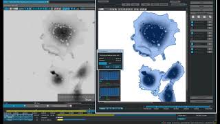
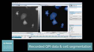
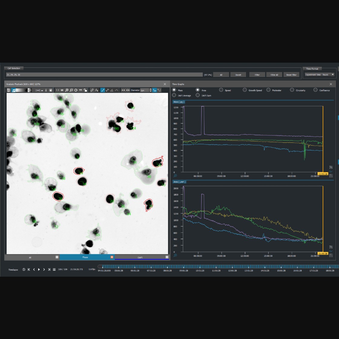
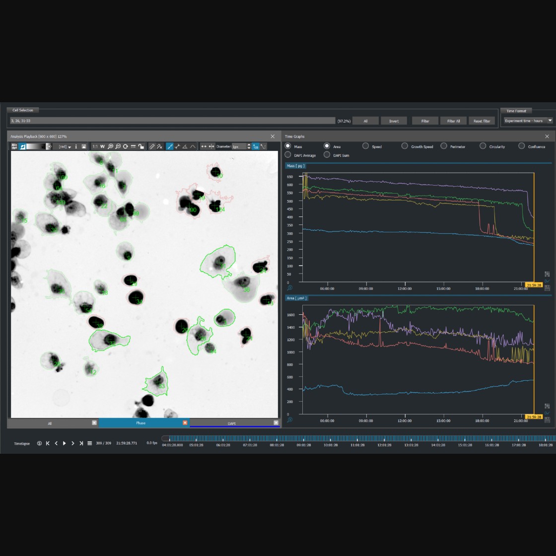
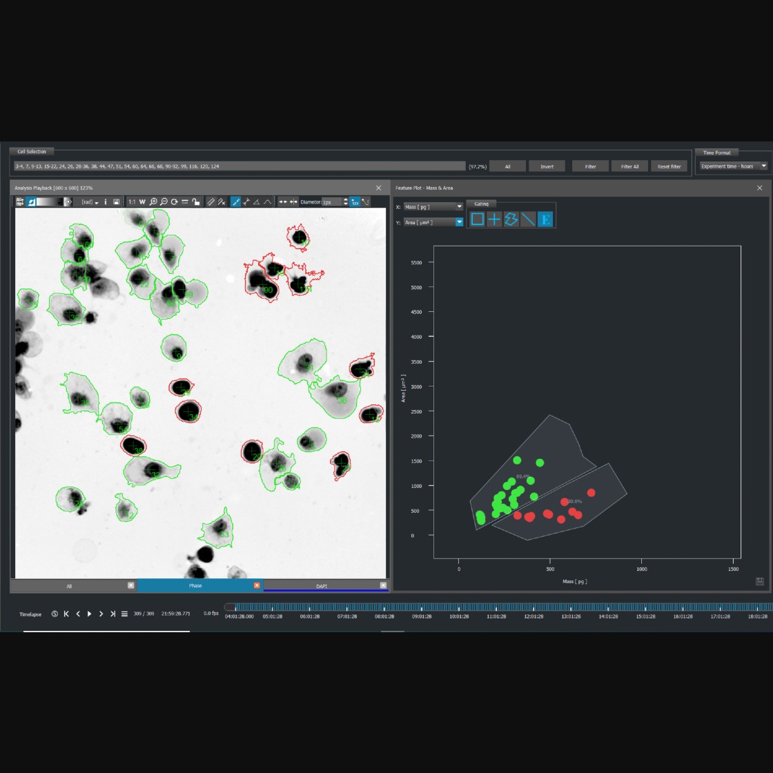
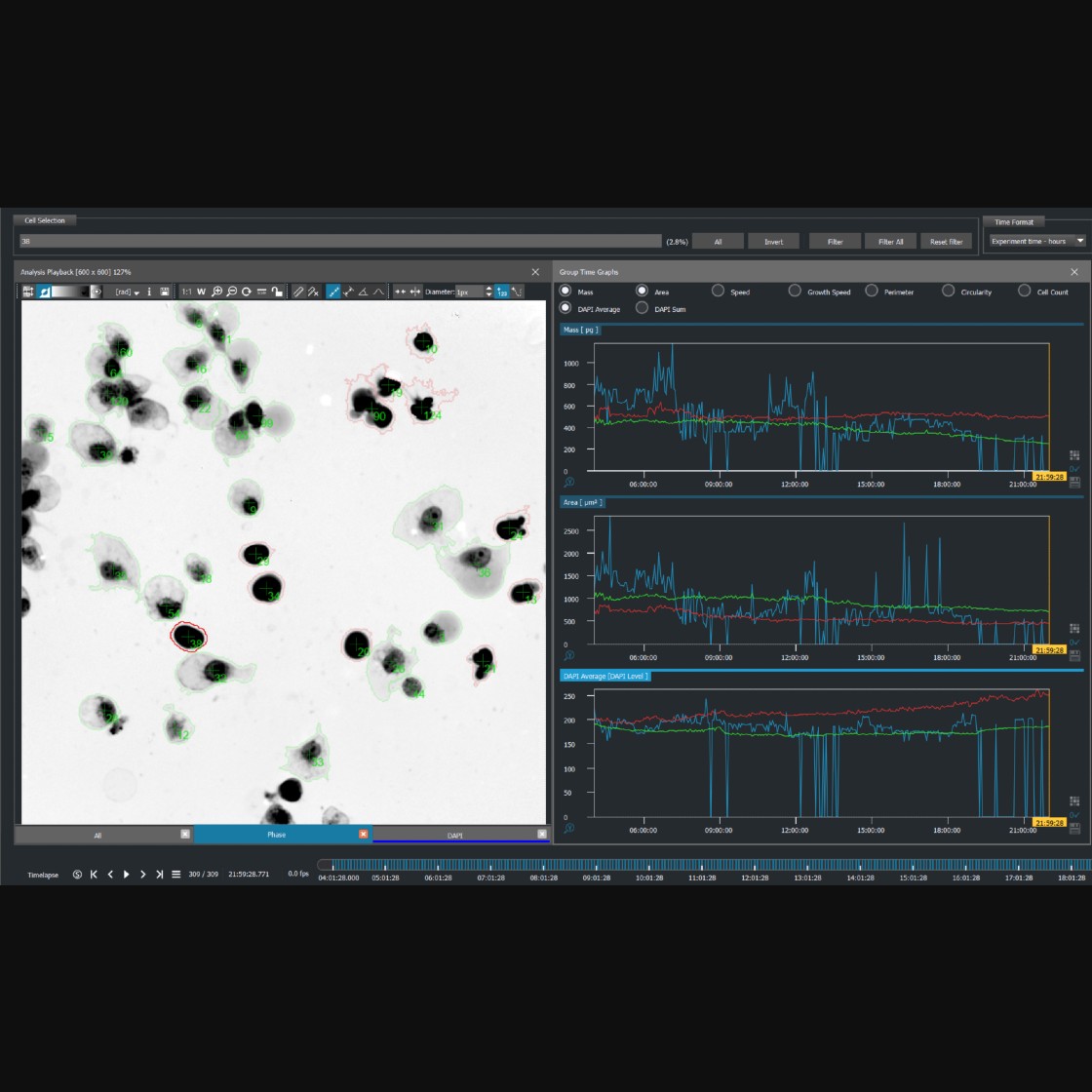
Q-Phase image quality permits very accurate and fast cell segmentation enabling to sort of the cells into distinct cell populations based on morphology, cell mass, or dynamics. A complete analysis of your experiment can be processed using All output data can be engaged using Q-Phase customized software.
Publications
Vicar, T. et al.
Cancer Cells Viscoelasticity Measurement by Quantitative Phase and Flow Stress Induction
Vicar, T. et al.
Self-Supervised Pretraining for Transferable Quantitative Phase Image Cell Segmentation
L. Štrbková, et al.
Classification of cells in time-lapse quantitative phase image by supervised machine learning
Cell segmentation methods for label-free contrast microscopy: review and comprehensive comparison
Products
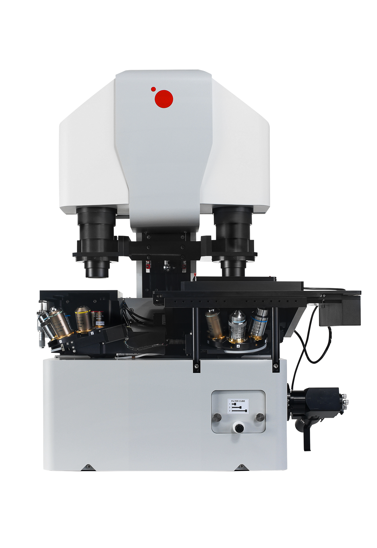
Telight Q-Phase
Quantitative Phase Imaging
Q-Phase is a patented holographic microscope with high detection sensitivity, designed for gentle live-cell imaging.
Q-Phase is an ideal solution for experts who desire precise automated segmentation of individual cells for subsequent data analysis. Q-Phase quickly transforms cell features and dynamics into numerical data suitable for comparisons, correlations, and more detailed statistics.
